The location of cartilage cells. Cartilage functions
 Live Journal
 Facebook
 Twitter
 Location of cartilage in the body n Cartilage tissues perform a form-forming function in the fetus and a supporting one in an adult body. Cartilaginous tissue can be found: n in the area of \u200b\u200bthe joints (covering the articular surface with a relatively narrow layer), n in the metaphyses (that is, between the pineal gland and the diaphysis) of the tubular bones, n in the intervertebral discs, in the anterior ribs, in the wall of the respiratory organs (larynx , trachea, bronchi), etc.
 Development n Like all other tissues of the internal environment of the body, skeletal tissues develop n from the mesenchyme (whose cells, in turn, are evicted from somites and splanchnotomes
 Features n The special nature of the intercellular substance gives two important properties: n elasticity and n strength. n of the intercellular substance of these tissues. n In many cases, the cartilage is covered by the perichondrium, a fibrous connective tissue that is involved in the growth and nutrition of cartilage.
 An important feature of cartilaginous tissue is the absence of blood vessels. Therefore, nutrients enter the cartilage - by diffusion from the vessels of the perichondrium.In some cases, there is no perichondrium - for example, in the articular cartilage, since their surface should be smooth. Here, nutrition is provided from the side of the synovial fluid and from the side of the underlying bone.
 Cellular composition n Chondroblasts - young cells, are located in the deep layers of the perichondrium one by one and are located closer to the surface of the cartilage n -small flattened cells capable of -proliferation and -synthesis of components of the intercellular substance of the cartilage. n they have well-expressed granular EPS, Golgi complex, mitochondria n Chondroblasts, releasing the components of the intercellular substance, "walled up" themselves in it and turn into chondrocytes.
 Functions n The main function of chondroblasts is the production of the organic part of the intercellular substance: collagen and elastin proteins, glycosaminoglycans (GAG) and proteoglycans (PG). n Chondroblasts provide appositional (superficial) growth of cartilage from the side of the perichondrium.
 Chondrocytes n a) Chondrocytes are the main type of cartilage cells. n - lie in special cavities of the intercellular substance (lacunae) and n - can divide by mitosis, while the daughter cells do not diverge, remain together - isogenic groups are formed (of 2-6 cells) originating from one cell. n b) They have n -large (compared to chondroblasts) size and oval shape. n Well-developed granular EPS and Golgi complex
 Functions n Chondrocytes that have stopped dividing actively synthesize the components of the intercellular substance. n Due to the activity of chondrocytes, there is an increase in the mass of cartilage from the inside - interstitial growth.
 Chondroclasts n In addition to the cells that form the intercellular substance, cartilage tissue also contains their antagonists - the destroyers of the intercellular substance - these are chondroclasts (can be attributed to the macrophagic system): rather large cells, there are many lysosomes and mitochondria in the cytoplasm. Function - destruction of damaged or worn out areas of cartilage.
 Intercellular substance n The intercellular substance of cartilage tissue contains fibers and basic substance. n many fibrous structures: n-collagen fibers, n and in elastic cartilage - elastic fibers.
 n The intercellular substance is highly hydrophilic, the water content reaches 75% of the mass of the cartilage, which determines the high density and turgor of the cartilage. Cartilage tissue in deep layers does not have blood vessels,
 n The main amorphous substance contains: n -water (70 -80%), -mineral substances (4 -7%), -organic component (10 -15%), represented by n -proteoglycans and -glycoproteins.
 Proteoglycans n Proteoglycan aggregate contains 4 components. n The aggregate is based on a long thread of hyaluronic acid (1). n With the help of globular binding proteins (2), n linear (fibrillar) peptide chains of the so-called crustal (core) protein (3). n In turn, oligosaccharide branches extend from the latter (4).
 These n complexes are highly hydrophilic; therefore, they bind a large amount of water and n provide high elasticity of the cartilage. n At the same time, they remain permeable to low molecular weight metabolites.
 n The perichondrium is the layer of connective tissue that covers the surface of the cartilage. In the perichondrium, the outer fibrous layer is isolated (from dense unformed CT with a large number of blood vessels) and the inner cell layer, which contains a large number of brain, semi-stem cells.
 Hyaline cartilage n Outwardly, this tissue is bluish-white in color and looks like glass (Greek hyalos - glass). Hyaline cartilage - covers all articular surfaces of bones, is contained in the sternal ends of the ribs, in the airways.
 Distinctive features of n 1. The intercellular substance of hyaline cartilage in preparations stained with hematoxylin-eosin seems homogeneous, without fibers. n 2. There is a clearly defined basophilic zone around isogenic groups - the so-called territorial matrix. This is due to the fact that chondrocytes secrete a large amount of GAGs with an acidic reaction, therefore this area is colored with basic paints, i.e., basophilic. Weakly oxyphilic areas between territorial matrices are called interterritorial matrix. n
 n A large number of proteoglycan aggregates. n Glycosaminoglycans. High elasticity depends on the content of GAG n Chondroitin sulfates (chondroitin-6-sulfate, chondroitin-4-sulfate) n Keratan sulfates n contains collagen type II, which is more hydrophilic (due to the higher content of hydroxy groups) and n forms only fibrils (not fiber). n Collagen IX, VI and X n Chondronectin protein
 Cellular composition n a) Immediately under the perichondrium there are n young chondrocytes (3) - n are somewhat larger in size and more oval in shape. n b) Deeper are n mature chondrocytes n large oval cells with light cytoplasm, n forming isogenic groups (4) of 2-6 cells.
 n 1) Joint surfaces of bones. n 2) Airways. n 3) The junction of the ribs with the sternum.
 Elastic cartilage n In the auricle, epiglottis, laryngeal cartilage. In the intercellular substance, in addition to collagen fibers, there is a large number of randomly located elastic fibers, which gives elasticity to the cartilage. Elastic cartilage has a lower content of lipids, chondroitin sulfates and glycogen.
 n b) in the thickness of the cartilaginous plate - isogenic groups of chondrocytes, n are large, oval and n have a light cytoplasm. n Groups of chondrocytes usually have n-type chains (of 2, rarely more cells), oriented perpendicular to the surface.
 Age-related changes n Due to the relatively low content of collagen fibrils and the absence of collagen X, the deposition of calcium salts (calcification) does not occur in elastic cartilage n due to malnutrition.
 Fibrous cartilage n Fibrous cartilage is located where tendons attach to bones and cartilage, intervertebral discs. In structure, it occupies an intermediate position between the densely formed connective and cartilaginous tissue. n
 n In the intercellular substance, there are much more collagen fibers located in an orientation - they form thick bundles, clearly visible under a microscope. Chondrocytes often lie singly along the fibers, without forming isogenic groups. They have an elongated shape, a rod-shaped nucleus and a narrow rim of the cytoplasm.
 n At the periphery, fibrous cartilage gradually passes n into a densely formed connective collagen fibers of which acquire orientation and go from one vertebra to another. tissue, oblique n b) In the central part of the disc, the fibrous cartilage passes into the nucleus pulposus, which contains hyaline cartilage, collagen type II (in the form of fibrils)
 Cartilage regeneration n Hyaline - negligible. The perichondrium is mainly involved n Elastic - less prone to degeneration and does not calcify n Fibrous - weak regeneration, capable of calcifying

 Composition n Bone tissue is composed of cells and extracellular substance. n Differon of bone tissue includes n 1. stem and semi-stem (osteogenic) cells, n osteoblasts, n osteocytes n 2. osteoclasts.
 Osteoblasts n Osteoblasts are the most functionally active cellular elements of the diferon during osteohistogenesis. In an adult organism, the source of cells supporting the osteoblast population is the cells of the dispersed cambium in the osteogenic layer of the periosteum. Osteoblasts have a cubic or prismatic shape. The nucleus is located eccentrically. Osteoblasts are typical actively synthesizing and secreting cells, secretion is carried out by the entire cell surface. The cell has a well-developed granular endoplasmic reticulum, which fills almost the entire cytoplasm, many free ribosomes and polysomes,
 Functions n secrete type I collagen, alkaline phosphatase, osteocalcin, osteopontin, transforming growth factors, osteonectin, collagenase, etc. n Highly differentiated osteoblasts are characterized by a gradual decrease in the activity of alkaline phosphatase, osteocalcin, osteopontin and the absence of proliferative activity.
 n Role in the mineralization of the organic basis of the bone matrix. The mineralization of the bone matrix begins with the deposition of amorphous calcium phosphate. Calcium cations enter the extracellular matrix from the bloodstream, where they are bound to proteins. n In the presence of alkaline phosphatase, synthesized by osteoblasts, glycerophosphates in the intercellular substance are broken down to form a phosphate anion. An excess of the latter leads to a local increase in Ca and P to a level at which calcium phosphate precipitates. The overwhelming fraction of the bone mineral is in the form of hydroxyapatite crystals. Crystals are formed on the collagen fibers of the bone matrix. The latter have structural features that facilitate this process. The fact is that the molecules of the collagen precursor, tropocollagen, are packed into a fiber in such a way that a gap remains between the end of one and the beginning of the other, called the hole zone. It is in this zone that the bone mineral is initially deposited. Subsequently, crystals begin to grow in both directions, and the process covers the entire fiber.
 n An essential role in the mineralization of the synthesized organic bone matrix belongs to the matrix vesicles. Such vesicles are derivatives of the Golgi complex of osteoblasts, have a membrane structure and contain various enzymes necessary for mineralization reactions or their inhibition, as well as amorphous calcium phosphates. The matrix vesicles exit the cells into the extracellular space and release the products enclosed in them. The latter initiate mineralization processes.
 Osteocytes n In terms of quantitative composition, the most numerous cells of bone tissue. These are process cells that lie in bone cavities - lacunae. The cell diameter reaches 50 microns. The cytoplasm is weakly basophilic. Organoids are poorly developed (granular EPS, PC and mitochondria). Do not share. n Function: take part in the physiological regeneration of bone tissue, produce the organic part of the intercellular substance. n The thyroid hormone calcitonin has a stimulating effect on osteoblasts and osteocytes - the synthesis of the organic part of the intercellular substance increases and the deposition of calcium increases, while the concentration of calcium in the blood decreases.


 Osteoclasts n n n Specialized macrophages. Their diameter reaches up to 100 microns. The various compartments of osteoclasts are specialized for specific functions. the basal zone, in which the genetic apparatus of the cell is concentrated in the composition of numerous (5 - 20) nuclei. a light area in direct contact with the bone matrix. Thanks to it, the osteoclast along the entire perimeter adheres tightly to the bone, creating an isolated space between itself and the surface of the mineralized matrix. Osteoclast adhesion is provided by a number of receptors to matrix components, the main of which are vitronectin receptors. The selective permeability of this barrier makes it possible to create a specific microenvironment in the cell adhesion zone. the vesicular area contains lysosomes. Enzymes, acidic substances are transported through the membrane of the corrugated border, carbonic acid H 2 CO 3 is formed; carbonic acid dissolves calcium salts, dissolved calcium is washed out into the blood. carrying out demineralization and disorganization of the bone matrix, which leads to the formation of a resorption (erosion) lacuna of Hauschip.
 Osteoclasts n Osteoclasts have many nuclei and a large volume of cytoplasm; the zone of cytoplasm adjacent to the bone surface is called a corrugated border, there are many cytoplasmic outgrowths and lysosomes of function - destruction of fibers and amorphous bone substance
 n Thick collagen fibers, devoid of cementitious substance, create a "brush border" Lysosomal enzymes proteolysis of collagen and other matrix proteins. Proteolysis products are removed from osteoclastic lacunae by transcellular transport. In general, the process of reducing the river. H in the lacuna is carried out by two mechanisms: by exocytosis of the acidic content of vacuoles in the lacuna and due to the action of proton pumps - H + -ATPases localized in the membrane of the corrugated border. The sources of hydrogen ions are water and carbon dioxide, which are the result of mitochondrial oxidation reactions.
 Intercellular substance n 1. Inorganic part of the matrix Contains a significant part of calcium (35%) and phosphorus (50%) (phosphoric acid and carbonate calcium salts) mainly in the form of crystals of hydroxyapatite (Ca 10 (PO 4) 6 (OH) 2 · (3 · Ca (OH) 2), n and a little - in the amorphous state, a small amount of magnesium phosphate - make up 70% of the intercellular substance.In the plasma, inorganic phosphorus is contained in the form of the anions HPO 4 -2 and Н 2 РО 4 -2. Nn The ratio of the organic and inorganic parts of the intercellular substance depends on age: in children, the organic part is slightly more than 30%, and the inorganic part is less than 70%, therefore their bones are less strong, but more flexible (not brittle); in old age, on the contrary, the proportion the inorganic part increases, and the organic part decreases, so the bones become harder, but more brittle.- blood vessels are present:
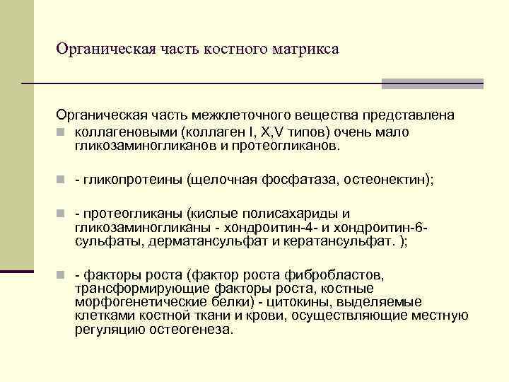 The organic part of the bone matrix The organic part of the intercellular substance is represented by n collagen (collagen I, X, V types), very few glycosaminoglycans and proteoglycans. n - glycoproteins (alkaline phosphatase, osteonectin); n - proteoglycans (acidic polysaccharides and glycosaminoglycans - chondroitin-4 - and chondroitin-6 sulfates, dermatan sulfate and keratan sulfate.); n - growth factors (fibroblast growth factor, transforming growth factors, bone morphogenetic proteins) - cytokines secreted by the cells of bone tissue and blood, carrying out local regulation of osteogenesis.
 proteins that carry out cell adhesion n Osteonectin is a glycoprotein of bone and dentin, has a high affinity for type I collagen and hydroxyapatite, and contains C-binding domains. Supports the concentration of Ca and P in the presence of collagen. It is assumed that the protein is involved in the interaction of the cell and the matrix. n Osteopontin is the main component of the protein composition of the matrix, in particular of the interfaces, where it accumulates in the form of a dense cover called cementation lines (lamina limitans). Due to its physicochemical properties, it regulates the calcification of the matrix, specifically participates in the adhesion of cells to the matrix or matrix to the matrix. Osteopontin production is one of the earliest manifestations of osteoblast activity. n Osteocalcin (OC) - a small protein (5800 Da, 49 amino acids) in the mineralized bone matrix, is involved in the process of calcification,
 Classification n Distinguish between tubular, flat and mixed bones. The diaphysis of tubular bones and cortical plates of flat and mixed bones are built of lamellar bone tissue covered with the periosteum or periosteum. In the periosteum, it is customary to distinguish between two layers: the outer layer is fibrous, consisting mainly of fibrous connective tissue; internal, adjacent to the surface of the bone - osteogenic, or cambial.
 Types of bone tissue coarse-fibrous (reticulofibrous) lamellar (fine-fibrous) Main feature Collagen fibers form a) Bone matter thick bundles running in different (organized into plates). directions. b) Moreover, within one plate, the fibers have the same direction, and within the adjacent plates, different. Localization 1. Flat bones of the embryo. 2. Bone tubercles; places of overgrown cranial sutures. Almost all bones of an adult are flat (scapula, pelvic bones, skull bones), spongy (ribs, sternum, vertebrae) and tubular.

 Lamellar bone tissue can be spongy and compact. Spongy bone substance Compact bone substance Localization Spongy substance consists of: epiphyses of tubular bones, inner layer (adjacent to the medullary canal) of diaphysis of tubular bones, spongy bones, inner part of flat bones. Most of the diaphysis of tubular bones and the surface layer of flat bones have a compact structure. Distinctive feature The spongy substance is built of avascular bone beams (beams), between which there are gaps - bone cells. There are practically no gaps in the compact bone substance: due to the growth of bone tissue deep into the cells, only narrow spaces for blood vessels remain - the so-called. central canals of osteons Bone marrow The cells of the spongy substance contain the vessels that feed the bone, and the red bone marrow, the hematopoietic organ. The bone marrow cavity of the diaphysis of tubular bones in adults contains yellow bone marrow - adipose tissue.
 Structure Consist of bone plates a) In this case, the plates of the spongy substance are usually oriented along the direction of the bone trabeculae, and not around the vessels, as in the osteons of the compact substance. b) osteons can occur in sufficiently thick beams. The unit of structure is bone plates. They consist of bone plates. In a compact substance, there are plates of 3 types: general (general) - they surround the entire bone, osteonic - lie in concentric layers around the vessel, forming the so-called. osteons; intercalated - are located between the osteons. osteons.
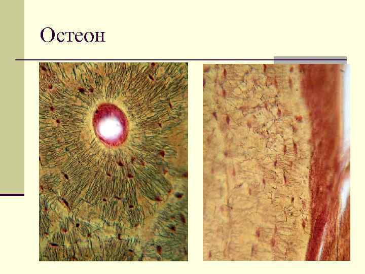
 The structure of the osteon, the main structural unit of the bone In the center of each osteon is a blood vessel (1), around the latter there are several concentric layers of bone plates (2), called osteonic ones. Osteons are delimited by a resorption (cleavage) line (3). Between the osteons there are intercalated bone plates (4), which are the remnants of the previous generations of osteons. bone plates include cells (osteocytes), collagen fibers and a basic substance rich in mineral compounds. the fibers in the intercellular substance are indistinguishable, and the intercellular substance itself has a solid consistency.
 Development of BONE FROM MESENCHYME (direct osteohistogenesis). From the mesenchyme, an immature (coarse-fibrous) bone is formed, which is subsequently replaced by a lamellar bone.In development, 4 stages are distinguished: n 1.the formation of an osteogenic islet - in the area of \u200b\u200bbone formation, mesenchymal cells turn into osteoblasts n
 2. the formation of the intercellular substance n osteoblasts begin to form the intercellular substance of the bone, while part of the osteoblasts is inside the intercellular substance, these osteoblasts turn into osteocytes; the other part of osteoblasts is on the surface of the intercellular substance,
 3. Calcification of the n intercellular substance of the bone The extracellular substance is impregnated with calcium salts. n a) At the third stage, the so-called. matrix vesicles similar to lysosomes. They store calcium and (due to alkaline phosphatase) inorganic phosphate. n b) At the rupture of bubbles, mineralization of the intercellular substance occurs, that is, the deposition of hydroxyapatite crystals on the fibers and in the amorphous substance. As a result, bone trabeculae (beams) are formed - mineralized areas of tissue containing all 3 types of bone cells - n n n from the surface - osteoblasts and osteoclasts, and in the depth - osteocytes.
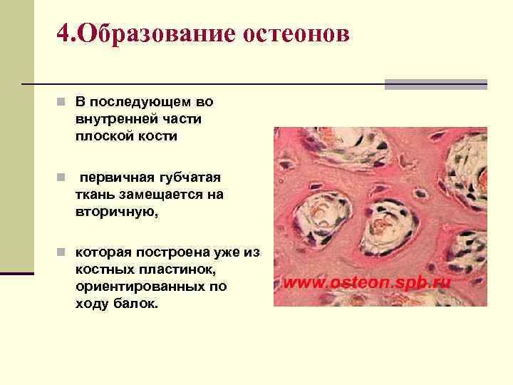 4. Formation of osteons n Subsequently, in the inner part of the flat bone n, the primary cancellous tissue is replaced by a secondary one, n which is already built of bone plates oriented along the beams.
 The development of lamellar bone tissue is closely related to 1. the process of destruction of individual sections of the bone and the ingrowth of blood vessels into the thickness of the reticulofibrous bone. Osteoclasts take part in this process both during the period of embryonic osteogenesis and after birth. 2. vessels growing to the trabeculae. In particular, around the vessels, the bone substance is formed in the form of concentric bone plates that make up the primary osteons.
 DEVELOPMENT OF BONE INTO THE SITE OF CARTELINE (indirect osteogenesis) n In place of cartilage, a mature (lamellar) bone is immediately formed n in development, there are 4 stages: n 1.cartilage formation - hyaline cartilage is formed at the site of the future bone
 2.perichondral ossification takes place only in the area of \u200b\u200bthe diaphysis in the area of \u200b\u200bthe diaphysis, the perichondrium turns into the periosteum, in which osteogenic cells appear, then osteoblasts due to osteogenic cells of the periosteum on the surface of the cartilage begin to form bone in the form of common plates with a circular course, like annual rings of a tree
 3. endochondral ossification n Occurs both in the diaphysis and in the pineal gland; blood vessels grow inside the cartilage, where there are osteogenic cells - osteoblasts, due to which bone in the form of osteons is formed around the vessels, and osteoclasts. n Cartilage breaks down simultaneously with bone formation
 bladder cartilage zone (4). At the border of the still preserved cartilage, cartilage cells are in a swollen, vacuolated state, i.e., the columnar cartilage zone has a vesicular shape (5). In the adjacent area of \u200b\u200bthe pineal gland, the growth of cartilage continues, and the multiplying cells line up in columns along the long axis of the bone.
 n a) Subsequently, ossification of the epiphysis itself (with the exception of the articular surface) will occur - by the enchondral route. n b) That is, mineralization will also occur here, n vessels will sprout here, the cartilage substance will collapse and first coarse-fibrous, n and then lamellar bone tissue is formed.
 n 4. bone restructuring and growth - old areas of bone are gradually destroyed and new ones are formed in their place; due to the periosteum, common bone plates are formed, due to osteogenic cells located in the adventitia of the bone vessels, osteons are formed. Between the diaphysis and the pineal gland there is a layer of cartilaginous tissue, due to which the growth of bone in length continues until the end of the period of growth of the body in length, that is, until 20-21 years.
 Bone growth Sources of growth Until the age of 20, tubular bones grow: in width - by appositional growth from the side of the perichondrium, in length - due to the activity of the metaepiphyseal cartilaginous plate. Metaepiphyseal cartilage a) The metaepiphyseal plate is a part of the pineal gland adjacent to the diaphysis and retaining (in contrast to the rest of the pineal gland) the cartilaginous structure. b) It has 3 zones (in the direction from the pineal gland to the diaphysis): borderline - contains oval chondrocytes, the zone of columnar cells - it is it that ensures the growth of cartilage in length due to the multiplication of chondrocytes, the zone of bladder cartilage - borders on the diaphysis and is ossified ... c) Thus, two processes occur simultaneously: the growth of cartilage (in the columnar zone) and its replacement by bone (in the vesicular zone).
 Regeneration n Regeneration and growth of bone in thickness is carried out due to the periosteum and endosteum. All tubular bones, as well as most of the flat bones, are histologically fine-fibrous bone.
 n In bone tissue, two oppositely directed processes constantly occur - resorption and neoplasm. The ratio of these processes depends on several factors, including age. The reconstruction of the bone tissue is carried out in accordance with the loads acting on the bone. n The process of bone tissue remodeling occurs in several phases, in each of which certain cells play a leading role. Initially, the site of bone tissue to be resorbed is "marked" by osteocytes using specific cytokines (activation). The protective layer on the bone matrix is \u200b\u200bdestroyed. Osteoclast precursors migrate to the bare bone surface, merge into a multinucleated structure - symplast - mature osteoclast. At the next stage, the osteoclast demineralizes the bone matrix (resorption), gives way to macrophages, which complete the destruction of the organic matrix of the intercellular substance of the bone and prepare the surface for osteoblast adhesion (reversion). At the last stage, precursors, differentiating into osteoblasts, arrive at the destruction zone, they synthesize and mineralize the matrix in accordance with the new conditions of static and dynamic load on the bone (formation).
In the human body, cartilaginous tissue serves as a support and connection between skeletal structures. There are several types of cartilaginous structures, each of which has its own location and performs its own tasks. Skeletal tissue undergoes pathological changes due to intense physical exertion, congenital abnormalities, age and other factors. To protect yourself from injuries and diseases, you need to take vitamins, calcium supplements and not get hurt.
The importance of cartilage structures
Articular cartilage holds the skeletal bones, ligaments, muscles and tendons together into a single musculoskeletal system. It is this type of connective tissue that provides shock absorption during movement, protecting the spine from damage, preventing fractures and bruises. The function of cartilage is to make the skeleton firm, elastic and flexible. In addition, cartilage forms a support frame for many organs, protecting them from mechanical damage.
Features of the structure of cartilage tissue
The specific gravity of the matrix exceeds the total weight of all cells. The general plan of the cartilage structure consists of 2 key elements: intercellular substance and cells. During the histological examination of the sample under the lenses of the microscope, the cells are located in a relatively smaller percentage of the area of \u200b\u200bspace. The intercellular substance contains about 80% water in the composition. The structure of the hyaline cartilage provides its main role in the growth and movement of the joints.
Intercellular substance
 Cartilage strength is determined by its structure.
 The matrix, as an organ of cartilage tissue, is heterogeneous and contains up to 60% of amorphous mass and 40% of chondrin fibers. Fibrils histologically resemble human skin collagen, but differ in a more chaotic distribution. The main substance of the cartilage consists of protein complexes, glucosaminoglycans, hyaluronan compounds and mucopolysaccharides. These components provide strong properties to the cartilage tissue, keeping it permeable to essential nutrients. There is a capsule, its name is perichondrium, it is a source of elements of cartilage regeneration.
Cellular composition
Chondrocytes are located in the intercellular substance rather chaotically. The classification divides cells into undifferentiated chondroblasts and mature chondrocytes. The precursors are formed by the perichondrium, and as they move into the deeper balls of tissue, the cells differentiate. Chondroblasts produce matrix ingredients, which include proteins, proteoglycans, and glucosaminoglycans. Young cells by dividing provide for the interstitial growth of cartilage.
Chondrocytes, located in the deep balls of tissue, are grouped into 3-9 cells, known as "isogenic groups". This mature cell type has a small nucleus. They do not divide, and their metabolic rate is greatly reduced. The isogenic group is encompassed by intertwined collagen fibers. The cells in this capsule are separated by protein molecules and have multiple shapes.
With degenerative-dystrophic processes, multinucleated chondroclast cells appear, which destroy and absorb tissues.
The table presents the main differences in the structure of types of cartilage tissue:
| View | Features: |
|---|---|
| Hyaline | Fine collagen fibers |
| Has basophilic and oxyphilic zones | |
| Elastic | Consists of elastin |
| Very flexible | |
| Has a honeycomb structure | |
| Fibrous | Formed from a large number of collagen fibrils |
| Chondrocytes are comparatively larger | |
| Lasting | |
| Capable of withstanding high pressure and compression |
Blood supply and nerves
 The tissue is not supplied with blood from its own vessels, but receives it by diffusion from nearby ones.
 Due to its very dense structure, cartilage does not have blood vessels of even the smallest diameter. Oxygen and all nutrients necessary for vital activity and functioning come by diffusion from nearby arteries, perichondrium or bone, and are also extracted from synovial fluid. Decomposition products are also removed diffusely.
In the upper balls of the perichondrium there are only a small number of individual branches of nerve fibers. Thus, the nerve impulse is not formed and does not propagate in pathologies. The localization of the pain syndrome is determined only when the disease destroys the bone, and the structures of the cartilage tissue in the joints are almost completely destroyed.
Varieties and functions
Depending on the type and position of fibrils, histology distinguishes the following types of cartilage tissue:
- hyaline;
- elastic;
- fibrous.
Each species is characterized by a certain level of elasticity, stability and density. The location of the cartilage determines its tasks. The main function of cartilage is to ensure the strength and stability of the joints between parts of the skeleton. The smooth hyaline cartilage found in the joints makes it possible for bones to move. Due to its appearance, it is called vitreous. Physiological conformity of the surfaces ensures smooth glide. The structural features of the hyaline cartilage and its thickness make it an integral part of the ribs, rings of the upper respiratory tract.
 The shape of the nose is formed by an elastic type of cartilage tissue.
 The elastic cartilage forms the appearance, voice, hearing and breathing. This refers to structures that are located in the framework of the small and medium-sized bronchi, auricles and the tip of the nose. The elements of the larynx are involved in the formation of a personal and unique timbre of the voice. Fibrous cartilage connects skeletal muscles, tendons, and ligaments to vitreous cartilage. Intervertebral and intra-articular discs and menisci are built from fibrous structures, they cover the temporomandibular and sternoclavicular joints.
The term "cartilage" refers to strong, elastic connective tissue. The human body has three types of cartilage: hyaline, elastic and bony. This article contains information to help you understand the types of cartilage in the human body.
Do you know that?
Hyaline cartilage forms most of the embryonic skeleton. Only after childbirth, the cartilage is replaced by bone tissue.
The term " connective tissue»Refers to a type of tissue that provides a supporting structure for the tissues and organs of the body. The properties of tissues are determined by the type of cells they contain, the number and location of fibers, and the properties of the basic substance (liquid part) from the matrix, which is located in the space between cells. Cartilage is a type of connective tissue that is formed from cells called chondrocytes. These cells can occur singly or in groups within gaps, which are gaps in the matrix. The perichondrium refers to a dense membrane, an irregular connective tissue that surrounds the surface of most cartilage tissue in the human body. It should be noted that the perichondrium is only the part of the cartilage where blood vessels and nerves can be found.
Even after the cartilage production process is complete, the chondrocytes remain inside the tissue. Then, they are called chondroblasts. In the case of cartilage, the base material is a gelatinous material called chondroitin sulfate. Collagen and elastin are protein fibers embedded in chondroitin sulfate. The matrix they form can be rigid or flexible. While the fibers in the matrix play a role in retention of shape and tensile strength, the hydrated, viscous matrix material protects the surrounding structures from compressive forces.
Most connective tissue has a rich blood supply, with the exception of cartilage. The delivery of nutrients through the blood is extremely important for quick recovery. Due to the minimal or limited supply of blood, cartilage injuries in adults take much longer to heal.
Types of cartilage tissue in the human body
There are three types of cartilage in the human body. These include:
- Hyaline
- Elastic
- Bone
These three types differ in terms of their elasticity, structure, strength, etc. Hyaline cartilage contains type II collagen fibers, which are widely dispersed, this elastic cartilage contains a large number of elastic fibers. Bony is the toughest of these and is densely packed with collagen fibers.
Hyaline cartilage is the most common type of cartilage. It has a pearly bluish color. Although tough and firm, it is also elastic.
It can be found in many places, including:
- Just below the thyroid cartilage, there is an annular part of the hyaline cartilage called cricoid cartilage.
- Where the ribs attach to the sternum
- In the trachea (in the form of tracheal rings, arytenoid cartilage (a pair of pyramidal cartilages) and cuneiform cartilage)
- In the primary bronchi, cartilage rings
- In the middle bronchi, like irregular cartilage plates
- Between joints such as knees, hips, shoulders, etc., such as articular cartilage

Articular cartilage covers the surface of the ends of the bones. It acts as a shock absorber. For example, the cartilage in the knees helps transfer stress when we run, sit, hang, or do any physical activity. The outer layer of cartilage is called the slip zone. One of the main functions of this type of cartilage is for the bones in the joints to move and slide over each other without friction. Based on the avascular position (lack of blood supply), the articular cartilage can become damaged due to natural wear and tear that occurs with age or injury. Degeneration of cartilage tissue in old age leads to a degenerative joint condition called arthrosis.
Elastic cartilage
Also known as yellow cartilage, this cartilage is quite elastic due to the presence of many irregular protein fibers in the matrix. The elastic fibers present in it are responsible for its ability to spring into shape immediately after it is deformed. It resembles hyaline cartilage to some extent.
The differentiating factor between the two types of cartilage is the presence of elastin fibers, which are embedded in the base material. The perichondrium is also found around this type of cartilage. It helps in shaping and maintaining the figure of certain structures of the body. It is a supportive, elastic fabric that provides firmness and flexibility.
It can be found in the following locations:
- The pinna or cartilaginous structure in the outer ear
- Eustachian tube
- Parts of the nose
- Parts of the larynx
- Parts of the epiglottis, which is the valve that closes the opening of the larynx when swallowing
The toughest type of cartilage is fibrous or bony cartilage, sometimes also called white. It has the ability to support heavy weights. The defining factor when it comes to histology is that it contains thin collagen fibers scattered in rows or layers. The number of chondrocytes is quite small, and the cells are embedded in the matrix material between the fibers, and not on the fibers. Bony contains both type I and type II collagen. This cartilage is very effective as a shock absorber due to its ability to withstand compressive forces. It provides support for surrounding structures attached to it.
It is found in the following locations in the human body:
- In the intervertebral discs
- On the knees
- Where the pelvic bones meet at the front of the body

Cartilage location
All three types of cartilage tissue are present in the human body and play an important role, especially the articular hyaline cartilage, which allows the joints to move freely. The other two types are also important, as they act as a cushion for a specific bone, provide support for surrounding structures, and withstand compressive forces. They can be damaged by injury or worn out with age. Displacement of the intervertebral disc and arthrosis are examples of damage to elastic, bone and hyaline cartilage, respectively. Since cartilage does not have a blood supply, its rate of recovery is rather slow when cartilage is damaged in adults. Therefore, you should make an appointment with your doctor as soon as possible for proper treatment.
3. Bone structure
4. Osteohistogenesis
1. Skeletal connective tissues include cartilaginous and bone tissues that perform supporting, protective and mechanical functions, as well as taking part in the metabolism of mineral substances in the body.
Cartilage tissue consists of cells - chondrocytes, chondroblasts and dense intercellular substance, consisting of amorphous and fibrous components. Chondroblasts are located singly along the periphery of the cartilaginous tissue. They are elongated flattened cells with a basophilic cytoplasm containing a well-developed granular endoplasmic reticulum and the Golgi apparatus. These cells synthesize the components of the intercellular substance, release them into the intercellular environment and gradually differentiate into definitive cells of the cartilage tissue - chondrocytes. Chondroblasts are capable of mitotic division. The perichondrium surrounding the cartilaginous tissue contains inactive, poorly differentiated forms of chondroblasts, which, under certain conditions, differentiate into chondroblasts, synthesizing the intercellular substance, and then into chondrocytes.
Chondrocytes by degree of maturity, according to morphology and function, they are subdivided into type I, II and III cells. All types of chondrocytes are localized in the deeper layers of cartilage tissue in special cavities - lacunae... Young chondrocytes (type I) divide mitotically, but daughter cells find themselves in one lacuna and form a group of cells - an isogenic group. The isogenic group is a common structural and functional unit of cartilage tissue. The location of chondrocytes in isogenic groups in different cartilage tissues is not the same.
Intercellular substance cartilage tissue consists of a fibrous component (collagen or elastic fibers) and an amorphous substance, which contains mainly sulfated glycosaminoglycans (primarily chondroitin sulfuric acids), as well as proteoglycans. Glycosaminoglycans bind large amounts of water and determine the density of the intercellular substance. In addition, the amorphous substance contains a significant amount of mineral substances that do not form crystals. Vessels in the cartilaginous tissue are normally absent.
Depending on the structure of the intercellular substance, cartilaginous tissues are divided into hyaline, elastic and fibrous cartilage tissue.
Hyaline cartilage characterized by the presence of only collagen fibers in the intercellular substance. In this case, the refractive index of the fibers and the amorphous substance is the same and therefore fibers in the intercellular substance are not visible on histological preparations. This also explains the certain transparency of the cartilage, consisting of hyaline cartilage tissue. Chondrocytes in isogenic groups of hyaline cartilage tissue are arranged in the form of rosettes. According to its physical properties, hyaline cartilage tissue is characterized by transparency, density and low elasticity. In the human body, hyaline cartilage tissue is widespread and is part of the large cartilage of the larynx (thyroid and cricoid), trachea and large bronchi, makes up the cartilaginous parts of the ribs, covers the articular surfaces of the bones. In addition, almost all bones of the body pass through the stage of hyaline cartilage during their development.
Elastic cartilage tissue characterized by the presence in the intercellular substance of both collagen and elastic fibers. In this case, the refractive index of elastic fibers differs from the refraction of an amorphous substance and therefore elastic fibers are clearly visible in histological preparations. Chondrocytes in isogenic groups in elastic tissue are arranged in columns or columns. According to its physical properties, elastic cartilage tissue is opaque, elastic, less dense and less transparent than hyaline cartilage tissue. It is part of elastic cartilage: auricle and cartilaginous part of the external auditory canal, cartilage of the external nose, small cartilage of the larynx and middle bronchi, and also forms the basis of the epiglottis.
Fibrous cartilage characterized by the content in the intercellular substance of powerful bundles of parallel collagen fibers. In this case, chondrocytes are located between the fiber bundles in the form of chains. Its physical properties are characterized by high strength. It occurs in the body only in limited places: it forms part of the intervertebral discs (annulus fibrosus), and is also localized at the points of attachment of ligaments and tendons to hyaline cartilage. In these cases, a gradual transition of connective tissue fibrocytes to cartilage chondrocytes is clearly traced.
There are two concepts that should not be confused - cartilage tissue and cartilage. Cartilage tissue - This is a type of connective tissue, the structure of which is described above. Cartilageis an anatomical organ that consists of cartilage tissue and perichondrium... The perichondrium covers the cartilaginous tissue from the outside (with the exception of the cartilage tissue of the articular surfaces) and consists of fibrous connective tissue.
Two layers are distinguished in the perichondrium:
external - fibrous;
internal - cellular or cambial (germ).
In the inner layer, poorly differentiated cells are localized - prechondroblasts and inactive chondroblasts, which, in the process of embryonic and regenerative histogenesis, transform first into chondroblasts and then into chondrocytes. The fibrous layer contains a network of blood vessels. Consequently, the perichondrium, as an integral part of the cartilage, performs the following functions: provides the avascular cartilage tissue with trophism; protects cartilage tissue; ensures the regeneration of cartilage tissue when damaged.
The trophism of the hyaline cartilage tissue of the articular surfaces is provided by the synovial fluid of the joints, as well as from the vessels of the bone tissue.
Development cartilage tissue and cartilage (chondrohistogenesis) is carried out from the mesenchyme. Initially, the mesenchymal cells in the places where the cartilaginous tissue is laid down intensively proliferate, round up and form focal cell clusters - chondrogenic islets... Then these rounded cells differentiate into chondroblasts, synthesize and secrete fibrillar proteins into the intercellular environment. Then chondroblasts differentiate into type I chondrocytes, which synthesize and secrete not only proteins, but also glycosoaminoglycans and proteoglycans, that is, they form the intercellular substance. The next stage in the development of cartilage tissue is the stage of differentiation of chondrocytes, while chondrocytes of type II, III appear and lacunae are formed. The perichondrium is formed from the mesenchyme surrounding the cartilaginous islets. In the process of cartilage development, two types of cartilage growth are noted: interstitial growth - due to the multiplication of chondrocytes and their release of the intercellular substance; oppositional growth - due to the activity of chondroblasts of the perichondrium and the imposition of cartilaginous tissue along the periphery of the cartilage.
Age-related changes are more marked in the hyaline cartilage tissue. In old and senile age, the deposition of calcium salts is noted in the deep layers of hyaline cartilage (cartilage mistletoe), germination of vessels into this area, and then replacement of calcified cartilaginous tissue with bone tissue - ossification... Elastic cartilage tissue is not subject to calcification and ossification, however, the elasticity of cartilage also decreases in old age.
2. Bone tissue is a type of connective tissue and consists of cells and intercellular substance, which contains a large amount of mineral salts, mainly calcium phosphate. Mineral substances make up 70% of the bone tissue, organic - 30%.
Bone tissue functions:
mechanical;
protective;
participation in the mineral metabolism of the body - a depot of calcium and phosphorus.
Bone cells: osteoblasts, osteocytes, osteoclasts. The main cells in the formed bone tissue are osteocytes... These are process cells with a large nucleus and weakly expressed cytoplasm (nuclear type cells). Cell bodies are localized in bone cavities - lacunae, and processes - in bone tubules. Numerous bone tubules, anastomosing each other, penetrate the entire bone tissue, communicating with the perivascular spaces, and form drainage system bone tissue. This drainage system contains tissue fluid, through which the exchange of substances is ensured not only between cells and tissue fluid, but also between the intercellular substance. The ultrastructural organization of osteocytes is characterized by the presence in the cytoplasm of a weakly expressed granular endoplasmic reticulum, a small number of mitochondria and lysosomes, and no centrioles. Heterochromatin predominates in the nucleus. All these data indicate that osteocytes have insignificant functional activity, which consists in maintaining the metabolism between cells and the intercellular substance. Osteocytes are definitive forms of cells and do not divide. They are formed from osteoblasts.
Osteoblasts contained only in developing bone tissue. They are absent in the formed bone tissue, but they are usually contained in an inactive form in the periosteum. In the developing bone tissue, they cover each bone plate along the periphery, tightly adhering to each other, forming a semblance of an epithelial layer. The shape of such actively functioning cells can be cubic, prismatic, angular. The cytoplasm of osteoblasts contains a well-developed granular endoplasmic reticulum and the lamellar Golgi complex, and many mitochondria. This ultrastructural organization suggests that these cells are synthesizing and secreting. Indeed, osteoblasts synthesize collagen protein and glycosaminoglycans, which are then secreted into the intercellular space. Due to these components, an organic matrix of bone tissue is formed. Then the same cells provide the mineralization of the intercellular substance through the release of calcium salts. Gradually, releasing the intercellular substance, they seem to be walled up and turn into osteocytes. At the same time, intracellular organelles are largely reduced, synthetic and secretory activity decreases, and the functional activity inherent in osteocytes remains. Osteoblasts, localized in the cambial layer of the periosteum, are in an inactive state, synthetic and transport organelles are poorly developed. When these cells are irritated (in the case of injuries, bone fractures, etc.), a granular endoplasmic reticulum and a lamellar complex rapidly develop in the cytoplasm, an active synthesis and release of collagen and glycosaminoglycans occurs, and the formation of an organic matrix (callus), and then the formation of definitive bone tissue. In this way, due to the activity of osteoblasts in the periosteum, bone regeneration occurs when they are damaged.
Oteoclasts - there are no bone-destroying cells in the formed bone tissue. But they are contained in the periosteum and in places of destruction and restructuring of bone tissue. Since the local processes of bone tissue restructuring are continuously carried out in ontogenesis, osteoclasts are necessarily present in these places. In the process of embryonic osteohistogenesis, these cells play an important role and are determined in large numbers. Osteoclasts have a characteristic morphology: firstly, these cells are multinucleated (3-5 or more nuclei), secondly, they are rather large cells (about 90 microns in diameter), and thirdly, they have a characteristic shape - the cell has an oval shape , but the part of it adjacent to the bone tissue is flat. At the same time, two zones are distinguished in the flat part:
the central part - corrugated contains numerous folds and islands;
the peripheral (transparent) part is in close contact with the bone tissue.
In the cytoplasm of the cell, under the nuclei, there are numerous lysosomes and vacuoles of various sizes. The functional activity of the osteoclast is manifested as follows: carbonic acid and proteolytic enzymes are released from the cytoplasm in the central (corrugated) zone of the cell base. Released carbonic acid causes demineralization of bone tissue, and proteolytic enzymes destroy the organic matrix of the intercellular substance. Fragments of collagen fibers are phagocytosed by osteoclasts and are destroyed intracellularly. Through these mechanisms, resorption(destruction) of bone tissue and therefore osteoclasts are usually localized in the depressions of the bone tissue. After the destruction of bone tissue due to the activity of osteoblasts, which are evicted from the connective tissue of the vessels, new bone tissue is built.
Intercellular substance bone tissue consists of a basic substance and fibers, which contain calcium salts. Fibers consist of type I collagen and are folded into bundles that can be arranged in parallel (ordered) or disordered, on the basis of which a histological classification of bone tissues is built. The main substance of bone tissue, like other types of connective tissue, consists of glycosaminoglycans and proteoglycans, but the chemical composition of these substances is different. In particular, bone tissue contains less chondroitinsulfuric acids, but more citric and other acids, which form complexes with calcium salts. In the process of development of bone tissue, an organic matrix-base substance and collagen (ossein, collagen type II) fibers are first formed, and then calcium salts (mainly phosphate) are deposited in them. Calcium salts form crystals of hydroxyapatite, which are deposited both in the amorphous substance and in the fibers, but a small part of the salts is deposited amorphous. Providing strength to bones, calcium phosphate salts are simultaneously a depot of calcium and phosphorus in the body. Therefore, bone tissue takes part in mineral metabolism.
Bone classification
There are two types of bone tissue:
reticulofibrous (coarse fibrous);
lamellar (parallel to fibrous).
IN reticulofibrous bone tissue the bundles of collagen fibers are thick, convoluted and disordered. In the mineralized intercellular substance, osteocytes are randomly located in the lacunae. Lamellar bone tissue consists of bone plates, in which collagen fibers or their bundles are located parallel in each plate, but at right angles to the course of the fibers in adjacent plates. Osteocytes are located between the plates in the lacunae, while their processes pass through the plates in the tubules.
In the human body, bone tissue is almost exclusively lamellar. Reticulofibrous bone tissue occurs only as a stage in the development of some bones (parietal, frontal). In adults, they are located in the area of \u200b\u200battachment of the tendons to the bones, as well as at the site of the ossified sutures of the skull (sagittal suture of the scales of the frontal bone).
When studying bone tissue, the concepts of bone tissue and bone should be differentiated.
3. Boneis an anatomical organ, the main structural component of which is bone... Bone as an organ consists of the following elements:
bone;
periosteum;
bone marrow (red, yellow);
vessels and nerves.
Periosteum (periosteum) surrounds bone tissue along the periphery (with the exception of articular surfaces) and has a structure similar to the perichondrium. In the periosteum, the outer fibrous and inner cellular or cambial layers are isolated. The inner layer contains osteoblasts and osteoclasts. A pronounced vascular network is localized in the periosteum, from which small vessels penetrate into the bone tissue through the perforating channels. Red bone marrow is considered as an independent organ and belongs to the organs of hematopoiesis and immunogenesis.
Bone in formed bones it is presented only in a lamellar form, however, in different bones, in a different part of one bone, it has a different structure. In the flat bones and epiphyses of tubular bones, the bone plates form crossbars (trabeculae)that make up the cancellous bone. In the diaphysis of the tubular bones, the plates are adjacent to each other and form a compact substance. However, even in a compact substance, some plates form osteons, while other plates are common.
The structure of the diaphysis of the tubular bone
On the transverse section of the diaphysis of the tubular bone, there are next layers:
periosteum (periosteum);
outer layer of general or general plates;
osteon layer;
inner layer of general or general plates;
internal fibrous plate
External common plates are located under the periosteum in several layers, but without forming complete rings. Osteocytes are located between the plates in the lacunae. Through the outer plates, piercing channels pass through which piercing fibers and vessels penetrate from the periosteum into the bone tissue. With the help of perforating vessels in the bone tissue, trophism is provided, and the perforating fibers connect the periosteum to the bone tissue.
Osteon layer consists of two components: osteons and insertion plates between them. Osteon - is a structural unit of the compact substance of the tubular bone. Every osteon comprises:
5-20 concentrically layered plates;
osteon channel, in which vessels pass (arterioles, capillaries, venules).
Between channels of adjacent osteons there are anastomoses. Osteons make up the bulk of the bone tissue of the diaphysis of the tubular bone. They are located longitudinally along the tubular bone, respectively, the lines of force and gravity and provide the performance of the supporting function. When the direction of the lines of force changes as a result of a fracture or curvature of the bones, osteons that do not bear the load are destroyed by osteoclasts. However, such osteons are not completely destroyed, and part of the osteon bone plates along its length is preserved and such remaining parts of the osteons are called insert plates... During postnatal ontogenesis, bone tissue is constantly being restructured - some osteons are destroyed (resorbed), others are formed, and therefore there are always insertion plates between the osteons, like the remnants of previous osteons.
The inner layer common records has a structure similar to the external one, but it is less pronounced, and in the area of \u200b\u200bthe transition of the diaphysis to the epiphyses, the common plates continue into the trabeculae.
Endost - thin connective tissue plate, lining the cavity of the diaphysis canal. The layers in the endosteum are not clearly expressed, but among the cellular elements there are osteoblasts and osteoclasts.
Bone and cartilage tissue make up the human skeleton. These tissues are entrusted with a supporting function, along with this they protect the internal organs, organ systems from adverse factors. For the normal functioning of the human body, it is necessary that all naturally occurring cartilages are in anatomically correct places, so that the tissues are strong and regenerate as needed. Otherwise, a person is faced with many unpleasant diseases that lower the standard of living, or even make it impossible to move independently.
Features of the fabric
Tissue, like any other structural elements of the body, is formed from special cells. Cartilage cells in science are called diferons. This concept is complex, it includes several types of cells: stem, semi-stem, united within the framework of anatomy in a group of little specialized ones - this category is inherent in the ability to actively divide. Chondroblasts are also isolated, that is, those cells that can divide, but at the same time are able to produce intercellular connections. Finally, there are cells whose main task is to create an intermediate substance. Their specialized name is chondrocytes. In the composition of these cells there are not only fibers of cartilage tissue, the function of which is to ensure stability, but also the main substance, which scientists call amorphous. This compound is able to bind water, so that the cartilage tissue resists compression stresses. If all the cells of the joint are healthy, it will be elastic and strong.
In science, there are three types of cartilage tissue. For division into groups, the features of the intercellular connecting component are analyzed. It is customary to talk about the following categories:
- elastic;
- hyaline;
- fibrous.
And if in more detail?
As is known from anatomy, all types of cartilage have their own characteristic features. So, elastic tissue is distinguished by the specifics of the structure of the intercellular substance - it is characterized by a rather high concentration of collagen fibers. At the same time, such fabric is rich in amorphous substance. At the same time, this fabric has a high percentage of elastic fibers, which gave it its name. The functions of the elastic-type cartilage tissue are associated with this feature: providing elasticity, flexibility, persistent resistance to external influences. What more interesting anatomy can tell? Where is this type of cartilage located? Usually - in those organs that are naturally provided for bending. For example, the laryngeal cartilage, nose and ear shells, and the center of the bronchi are composed of elastic cartilage tissue.
Fibrous fabric: some features
At the point from which the hyaline cartilage begins, the fibrous connective tissue ends. Usually, this tissue is found in the discs between the vertebrae, as well as at the junction of bones, where mobility is not important. The structural features of this type of cartilage tissue are directly related to the specifics of its location. Tendons, ligaments at the point of contact with the cartilage tissue provoke an actively developed system of collagen fibers. The peculiarity of such tissue is the presence of cartilage cells (instead of fibroblasts). These cells form isogenic groups.

What else do you need to know
The course of human anatomy allows you to clearly understand what cartilage tissue is needed for: to ensure mobility while maintaining elasticity, stability, security. These fabrics are dense and provide mechanical protection. Modern anatomy as a science is characterized by an abundance of terms, including those complementary and mutually replacing each other. So, if we are talking about the vitreous cartilage tissue of the spine, then it is assumed that they are talking about hyaline. It is this tissue that forms the ends of the bones that make up the rib cage. Some elements of the respiratory system are also created from it.
The function of cartilage tissue from the category of connective tissue is the connection of tissue and hyaline vitreous cartilage, which has a completely different structure. But the reticular cartilage tissue ensures the normal functioning of the epiglottis, the hearing system, and the larynx.
Why is cartilage tissue needed?
Nature does not create anything for nothing. All tissues, cells, organs have a fairly extensive functionality (and some tasks are still hidden from scientists). As is already known from anatomy today, the functions of cartilage tissue include a guarantee of the reliability of the connection of elements that provide a person with the ability to move. In particular, the bony elements of the spine are connected with each other by cartilage tissue.
As it was possible to establish in the course of studies on aspects of nutrition of cartilage tissue, it takes an active part in carbohydrate metabolism. This explains some of the features of regeneration. It is noted that in childhood, the restoration of cartilaginous tissue is possible by 100%, but after years this ability is lost. If an adult is faced with damage to the cartilage tissue, he can only rely on partial restoration of mobility. At the same time, the restoration of cartilage tissue is one of the tasks attracting the attention of the leading medical minds of our time, therefore it is assumed that it will be possible to find an effective pharmaceutical solution to this problem in the near future.
Joint problems: there are options
Currently, medicine can offer several methods for restoring organs and tissues damaged for various reasons. If the joint has received a mechanical injury or a certain disease has provoked the destruction of biological material, in most cases, prosthetics becomes the most effective solution to the problem. But injections for the cartilage tissue will help when the situation has not gone so far, degenerative processes have begun, but are reversible (at least partially). As a rule, they resort to means that contain glucosamine, sodium sulfate.
Understanding how to restore cartilage tissue at the initial stages of the disease, they usually resort to physical exercise, strictly monitoring the level of stress. A good effect is shown by therapy using inflammation-blocking drugs. As a rule, most patients are prescribed medications that are rich in calcium in a form that is easily absorbed by the body.
Cartilaginous connective tissue: where do the problems come from?
In most cases, the disease is provoked by previous injuries or infection of the joint. Sometimes the degeneration of cartilaginous connective tissue is provoked by increased loads falling on it for a long time period. In some cases, problems are associated with genetic backgrounds. Hypothermia of body tissues can play a role.

In case of inflammation, the use of drugs for external use and tablets can give a good result. Modern medicines are formed taking into account the hydrophilicity characteristic of the cartilaginous tissue of the spine and other organs. This means that topical agents can quickly "get" to the affected area and have a therapeutic effect.
Structural features
As can be seen from the anatomy, hyaline cartilage, other cartilage tissues, as well as bone are grouped into the skeletal category. In Latin, this group of tissues is called textus cartilaginus. Up to 80% of this tissue is water, from four to seven percent is salt, and the rest is organic components (up to 15%). The dry part of cartilage tissue is half or more (up to 70%) formed from collagen. The matrix produced by tissue cells is a complex substance that includes hyaluronic acid, glycosaminoglycans, proteoglycans.
Tissue cells: some features
As scientists have found out, chondroblasts are young cells that usually have an irregular elongated shape. In the process of vital activity, such a cell generates proteoglycans, elastin, and other components essential for the normal functioning of the joint. The cytolemma of such a cell is a large number of microvilli. The cytoplasm contains an abundance of RNA. Such a cell is characterized by an endoplasmic reticulum of a high level of development, presented both in a non-granular form and in a granular one. The cytoplasm of chondroblasts also contains glycogen granules, the Golgi complex, and lysosomes. Usually the nucleus of such a cell has one or two nuclei. The formation contains a large amount of chromatin.
A distinctive feature of chondrocytes is their large size, since these cells are already mature. They are characterized by a round, oval, polygonal shape. Most chondrocytes are equipped with processes, organelles. Typically, such cells occupy lacunae, and around them is an intercellular connective substance. When a lacuna contains one cell, it is classified as primary. Isogenic groups consisting of a pair or three of cells are predominantly observed. This allows us to speak of a secondary lacuna. The wall of this formation has two layers: on the outside it is made of collagen fibers, and on the inside it is lined with proteoglycan aggregates interacting with the cartilaginous glycocalyx.
Biological features of tissue
When the cartilaginous tissue of the joint is in the focus of attention of scientists, it is usually studied as an accumulation of chondrons - this is the name given to the functional, structural units of biological tissue. A chondron is formed from a cell or a combined group of cells, a matrix surrounding the cell, and capsule-shaped lacunae. Each of the three types of cartilage listed above has its own unique structural features. For example, hyaline cartilage, which got its name from the Greek word for "glass", has a bluish tint and is characterized by cells of the most varied shapes and structures. Much depends on what place the cell occupies inside the cartilage tissue. Usually, hyaline cartilage is formed by groups of chondrocytes. Such tissue creates joints, rib cartilage, larynx.

If we consider the process of bone formation in the human body, you can see that at the initial stage, most of them consist of hyaline cartilage. Over time, the transformation of articular tissue into bone occurs.
What else is special?
But fibrous cartilage is very strong, as it consists of thick fibers. Its cells are characterized by an elongated shape, a rod-shaped nucleus and cytoplasm, which forms a small rim. Such cartilage usually creates fibrous rings characteristic of the spine, menisci, discs inside the joints. Cartilage covers some of the joints.
If we consider the elastic cartilage tissue, you will notice that it is quite flexible, since the matrix is \u200b\u200brich not only in collagen, but also in elastic fibers. This tissue is characterized by rounded cells enclosed in lacunae.
Cartilage and cartilage tissue
These two terms, despite their similarities, should not be confused. Cartilage is a type of connective biological tissue, while cartilage is an anatomical organ. In its structure, there is not only cartilage tissue, but also the perichondrium, which covers the tissues of the organ from the outside. In this case, the perichondrium does not cover the articular surface. This element of cartilage is formed by a connective tissue made up of fibers.

The perichondrium consists of two layers: fibrous, covering it from the outside, and cambial, with which the organ is lined inside. The second is also known as sprout. The inner layer is an accumulation of poorly differentiated cells. These include chondroblasts in an inactive stage, prechondroblasts. From these cells, chondroblasts are first formed, then they progress to chondrocytes. But the fibrous layer is distinguished by a developed circulatory network, represented by an abundance of blood vessels. The perichondrium is both a protective layer and a storage of material for regenerative processes, and a tissue, thanks to which the trophism of cartilaginous tissue is realized, in the structure of which there are no vessels. But if we consider hyaline cartilage, then in it the main tasks for trophism fall on the synovial fluid, and not only on the vessels. The blood supply to the bone tissue plays a very important role.
How it works?
The basis for the formation of cartilage, cartilage tissue is the mesenchyme. The process of tissue growth in science is called chondrogistogenesis. Mesenchymal cells at points where nature provides for the presence of cartilaginous tissue multiply, divide, grow, and round up. This results in a cell clump called a focus. Science usually refers to such places as chondrogenic islets. As the process moves forward, differentiation into chondroblasts occurs, due to which the production of fibrillar proteins that enter the environment between living cells becomes real. This leads to the formation of the first type of chondrocytes, capable of not only producing specialized proteins, but also a number of other compounds that are indispensable for the normal activity of organs.
As the cartilage tissue develops, chondrocytes differentiate, which leads to the formation of the second and third types of cells in this tissue. At the same stage, gaps appear. The mesenchyme, located around the cartilaginous islet, becomes the source of cells for the creation of the perichondrium.
Features of tissue growth
The development of cartilage is usually divided into two stages. First, the tissues go through a period of interstitial growth, during which chondrocytes actively multiply and produce intercellular substance. Then comes the stage of oppositional growth. Here "the main characters" are the chondroblasts of the perichondrium. In addition, tissue overlays located at the periphery of the organ provide irreplaceable assistance for the formation and functioning of cartilage tissue.

With the aging of the body as a whole, cartilaginous tissue in particular, degenerative processes are outlined. The most prone to such are hyaline cartilage. Elderly people are often faced with pain provoked by salt deposits in the deep cartilage layers. Calcium compounds accumulate more often, which leads to tissue blurring. The vessels grow into the affected area, the cartilage tissue is gradually transformed into bone. In medicine, this process is called ossification. But elastic tissues are not damaged by such changes, they do not ossify, although they lose elasticity over the years.
Cartilage tissue: degeneration problems
It so happened that from the point of view of human health, cartilage tissue is one of the most vulnerable, and almost all elderly people, and often the younger generation, suffer from diseases associated with joints. There are many reasons for this: it is the ecology, and the wrong way of life, and the wrong diet. Of course, very often we get injured, we face infections or inflammations. A one-time problem - injury or illness - passes, but at an older age it returns with echoes - joint pain.
Cartilage is quite sensitive to many diseases. Problems with the musculoskeletal system arise if a person is faced with a hernia, dysplasia, arthrosis, arthritis. Some suffer from a lack of natural collagen synthesis. With age, chondrocytes degenerate, and cartilage tissue suffers greatly from this. In many cases, the best therapeutic effect is provided by surgery, when the affected joint is replaced with an implant, but this solution is not always applicable. If there is a chance of restoring natural cartilage tissue, this chance should not be neglected.
Joint diseases: how do they manifest?
The majority of those suffering from such pathologies can more accurately predict the change in weather than any forecast: the joints affected by the disease respond to the slightest changes in the surrounding space with excruciating, pulling pain. If the patient suffers from joint damage, he cannot move abruptly, since the tissues react to this with sharp, severe pain. As soon as similar symptoms begin to appear, you need to immediately make an appointment with a doctor. It is much easier to cure a disease or block its development if you start the fight at an early stage. Delay leads to the fact that regeneration becomes completely impossible.
To restore the normal functionality of cartilage tissue, quite a few drugs have been developed. Mostly they are classified as non-steroidal and are designed to block inflammation. Also produced are pain relievers - pills, injections. Finally, recently, special chondroprotectors have become widespread.
How to treat?
The most effective remedies against degenerative processes in cartilage tissue affect the cellular level. They block inflammatory processes, protect chondrocytes from the negative effects, and also stop the degenerative activity of various aggressive compounds that attack cartilage tissue. Once the inflammation has been effectively blocked, the next step in therapy is usually to restore the intercellular junction. For this, chondroprotectors are used.
Several agents of this group have been developed - they are built on different active components, which means that they differ in the mechanism of action on the human body. For all funds in this group, effectiveness is characteristic only when taken in a long course, which allows you to achieve really good results. Particularly widespread are preparations made on chondroitin sulfate. It is glucosamine, which is involved in the formation of cartilage proteins and helps restore tissue structure. Due to the supply of a substance from an external source to all types of cartilage tissue, the process of production of collagen, hyalic acid is activated, and the cartilage is independently restored. With the correct use of medicines, you can quickly restore joint mobility and get rid of pain.

Another good option is products containing other glucosamines. They restore tissue from all sorts of damage. Under the influence of the active component, the metabolism in the cartilaginous tissues of the joint is normalized. Also recently used drugs of animal origin, that is, made from biological material obtained from animals. Most often these are the tissues of calves, aquatic creatures. Good results are shown by therapy using mucopolysaccharides and medicines based on them.
- Causes of Feeling Shaky and Unsteady When Walking
- Tertiary structure of proteins Native structure of protein and its disorders
- What is eps in biology
- Why you need to eat soup. Is the soup healthy? What should be the right soup
- Why, before death, do people see the dead, empty themselves, and their nose sharpens?
- Emergency care for coma algorithm of actions
- Neurological diseases: list, symptoms, causes and treatment features