Structural organization of proteins. Tertiary structure of proteins Native structure of protein and its disorders
 Live Journal
 Facebook
 Twitter
MODULE 1 STRUCTURE, PROPERTIES AND FUNCTIONS OF PROTEINS
MODULE 1 STRUCTURE, PROPERTIES AND FUNCTIONS OF PROTEINS
Module structure | Topics |
Modular unit 1 | 1.1. Structural organization of proteins. Stages of the formation of native protein conformation 1.2. Fundamentals of protein functioning. Drugs as ligands that affect protein function 1.3. Denaturation of proteins and the possibility of their spontaneous renewal |
Modular unit 2 | 1.4. Features of the structure and functioning of oligomeric proteins on the example of hemoglobin 1.5. Maintaining native conformation of proteins under cell conditions 1.6. Variety of proteins. Protein families as exemplified by immunoglobulins 1.7. Physicochemical properties of proteins and methods of their separation |
Modular unit 1 STRUCTURAL ORGANIZATION OF MONOMERIC PROTEINS AND THE BASIS OF THEIR FUNCTIONING
Learning Objectives To be able to:
1. Use knowledge about the features of the structure of proteins and the dependence of the functions of proteins on their structure to understand the mechanisms of development of hereditary and acquired proteinopathies.
2. Explain the mechanisms of the therapeutic action of some drugs as ligands that interact with proteins and change their activity.
3. Use knowledge of the structure and conformational lability of proteins to understand their structural and functional instability and tendency to denaturation under changing conditions.
4. Explain the use of denaturing agents as means for sterilizing medical material and instruments, as well as antiseptics.
Know:
1. Levels of the structural organization of proteins.
2. The value of the primary structure of proteins, which determines their structural and functional diversity.
3. The mechanism of formation of an active center in proteins and its specific interaction with the ligand, which underlies the functioning of proteins.
4. Examples of the influence of exogenous ligands (drugs, toxins, poisons) on the conformation and functional activity of proteins.
5. Causes and consequences of protein denaturation, factors causing denaturation.
6. Examples of the use of denaturing factors in medicine as antiseptics and means for sterilizing medical instruments.
TOPIC 1.1. STRUCTURAL ORGANIZATION OF PROTEINS. STAGES OF FORMATION OF NATIVE
PROTEIN CONFORMATIONS
Proteins are polymer molecules with a total of 20 α-amino acids as monomers. The set and order in which amino acids are combined in a protein is determined by the structure of genes in the DNA of individuals. Each protein, in accordance with its specific structure, performs its own function. The set of proteins of a given organism determines its phenotypic characteristics, as well as the presence of hereditary diseases or a predisposition to their development.
1. Amino acids that make up proteins. Peptide bond.Proteins are polymers built from monomers - 20 α-amino acids, the general formula of which is
Amino acids differ in structure, size, physicochemical properties of the radicals attached to the α-carbon atom. The functional groups of amino acids determine the characteristics of the properties of different α-amino acids. The radicals found in α-amino acids can be divided into several groups:
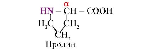
Proline,unlike other 19 protein monomers, it is not an amino acid, but an imino acid, the radical in proline is linked to both the α-carbon atom and the imino group
Amino acids differ in their solubility in water.This is due to the ability of radicals to interact with water (hydrate).
TO hydrophilicincludes radicals containing anionic, cationic and polar uncharged functional groups.
TO hydrophobicincludes radicals containing methyl groups, aliphatic chains or cycles.
2. Peptide bonds link amino acids into peptides.In the synthesis of a peptide, the α-carboxyl group of one amino acid interacts with the α-amino group of another amino acid to form peptide bond:
Proteins are polypeptides, i.e. linear polymers of α-amino acids linked by a peptide bond (Fig.1.1.)
 Figure: 1.1. Terms used to describe the structure of peptides
Monomers of amino acids that make up polypeptides are called amino acid residues.A chain of repeating groups - NH-CH-CO- forms peptide backbone.An amino acid residue that has a free α-amino group is called N-terminal, and one that has a free α-carboxyl group is called C-terminal. The peptides are written and read from the N-terminus to the C-terminus.
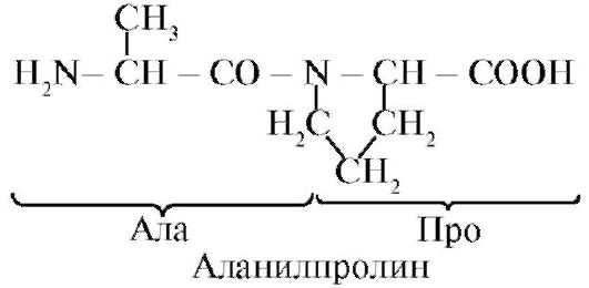
The peptide bond formed by the imino group of proline differs from other peptide bonds: the nitrogen atom of the peptide group lacks hydrogen,
instead, there is a bond with the radical, as a result, one side of the cycle is included in the peptide backbone:
 Peptides differ in amino acid composition, the number of amino acids and the order in which the amino acids are combined, for example, Ser-Ala-Glu-Gis and Gis-Glu-Ala-Ser are two different peptides.
Peptide bonds are very strong, and strict conditions are required for their chemical non-enzymatic hydrolysis: the analyzed protein is hydrolyzed in concentrated hydrochloric acid at a temperature of about 110 ° for 24 hours. In a living cell, peptide bonds can be broken using proteolytic enzymes,called proteasesor peptide hydrolases.
3. Primary structure of proteins.Amino acid residues in peptide chains of different proteins do not alternate randomly, but are arranged in a certain order. The linear sequence or the order of alternation of amino acid residues in the polypeptide chain is called the primary structure of the protein.
The primary structure of each individual protein is encoded in a DNA molecule (in a region called a genome) and is realized during transcription (rewriting information on mRNA) and translation (synthesis of the primary structure of a protein). Consequently, the primary structure of proteins of an individual person is information hereditarily transmitted from parents to children, which determines the structural features of the proteins of a given organism, on which the function of existing proteins depends (Fig. 1.2.).
 Figure: 1.2. The relationship between the genotype and the conformation of proteins synthesized in the body of an individual
Each of the approximately 100,000 individual proteins in the human body has uniqueprimary structure. The molecules of one type of protein (for example, albumin) have the same alternation of amino acid residues, which distinguishes albumin from any other individual protein.
The sequence of amino acid residues in the peptide chain can be considered as a form of recording information. This information determines the spatial folding of the linear peptide chain into a more compact three-dimensional structure called conformationsquirrel. The process of forming a functionally active protein conformation is called folding.
4. Conformation of proteins.Free rotation in the peptide backbone is possible between the nitrogen atom of the peptide group and the adjacent α-carbon atom, as well as between the α-carbon atom and the carbonyl carbon. Due to the interaction of functional groups of amino acid residues, the primary structure of proteins can acquire more complex spatial structures. In globular proteins, there are two main levels of folding of the conformation of peptide chains: secondaryand tertiary structure.
Secondary structure of proteinsis a spatial structure formed as a result of the formation of hydrogen bonds between the functional groups -C \u003d O and - NH- of the peptide backbone. In this case, the peptide chain can acquire regular structures of two types: α-helixand β-structures.
IN α-helixhydrogen bonds are formed between the oxygen atom of the carbonyl group and the hydrogen of the amide nitrogen of the 4th amino acid from it; side chains of amino acid residues
are located along the periphery of the spiral, without participating in the formation of the secondary structure (Fig. 1.3.).
Bulky radicals or radicals carrying the same charges prevent the formation of the α-helix. The proline residue, which has a ring structure, interrupts the α-helix, since a hydrogen bond cannot be formed due to the absence of hydrogen at the nitrogen atom in the peptide chain. The bond between nitrogen and the α-carbon atom is part of the proline cycle; therefore, the peptide backbone at this point acquires a bend.
β-structureforms between the linear regions of the peptide backbone of one polypeptide chain, thus forming folded structures. Polypeptide chains or parts thereof can form parallelor antiparallel β-structures.In the first case, the N- and C-ends of the interacting peptide chains coincide, and in the second, they have the opposite direction (Fig. 1.4).
Figure: 1.3. Secondary structure of protein - α-helix
 Figure: 1.4. Parallel and antiparallel β-fold structures
β-structures are indicated by wide arrows: A - Antiparallel β-structure. B - Parallel β-folded structures
In some proteins, β-structures can be formed due to the formation of hydrogen bonds between the atoms of the peptide backbone of different polypeptide chains.
Also found in proteins areas with irregular secondarystructure, which include bends, loops, turns of the polypeptide backbone. They are often located in places where the direction of the peptide chain changes, for example, when a parallel β-sheet structure is formed.
According to the presence of α-helices and β-structures, globular proteins can be divided into four categories.
Figure: 1.5. Secondary structure of myoglobin (A) and β-chains of hemoglobin (B), containing eight α-helices

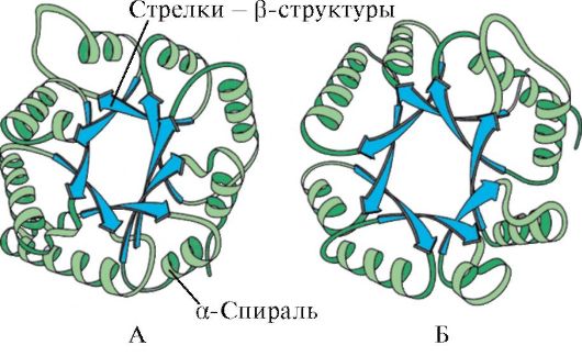 Figure: 1.6. Secondary structure of triose phosphate isomerase and pyruvate kinase domain
 Figure: 1.7. Secondary structure of the constant domain of immunoglobulin (A) and the enzyme superoxide dismutase (B)
IN fourth categoryincluded proteins containing a small amount of regular secondary structures. These proteins include small, cysteine-rich proteins or metalloproteins.
Protein tertiary structure- the type of conformation formed due to interactions between amino acid radicals, which can be located at a considerable distance from each other in the peptide chain. In this case, most proteins form a spatial structure resembling a globule (globular proteins).
Since hydrophobic amino acid radicals tend to combine with the so-called hydrophobic interactionsand intermolecular van der Waals forces, a dense hydrophobic core is formed inside the protein globule. Hydrophilic ionized and non-ionized radicals are mainly located on the surface of the protein and determine its solubility in water.
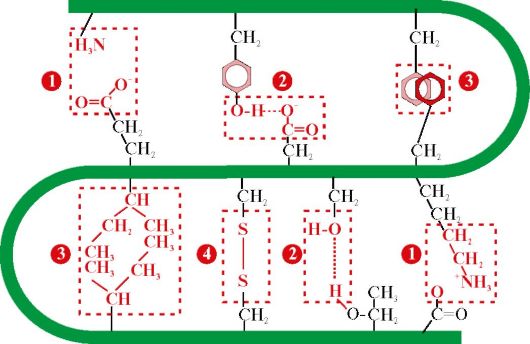 Figure: 1.8. Types of bonds that arise between amino acid radicals during the formation of the tertiary structure of a protein
1 - ionic bond- occurs between positively and negatively charged functional groups;
2 - hydrogen bond- occurs between an uncharged hydrophilic group and any other hydrophilic group;
3 - hydrophobic interactions- arise between hydrophobic radicals;
4 - disulfide bond- formed due to the oxidation of SH-groups of cysteine \u200b\u200bresidues and their interaction with each other
Hydrophilic amino acid residues trapped inside the hydrophobic core can interact with each other using ionicand hydrogen bonds(fig. 1.8).
Ionic and hydrogen bonds, as well as hydrophobic interactions, are among the weak ones: their energy is not much higher than the energy of the thermal motion of molecules at room temperature. Protein conformation is maintained through the emergence of many of these weak bonds. Since the atoms that make up the protein are in constant motion, some weak bonds can be broken and others formed, which leads to small displacements of individual sections of the polypeptide chain. This property of proteins to change conformation as a result of breaking some and the formation of other weak bonds is called conformational lability.
The human body has systems that support homeostasis- the constancy of the internal environment within certain limits permissible for a healthy organism. Under homeostasis conditions, small changes in conformation do not disrupt the overall structure and function of proteins. The functionally active conformation of a protein is called native conformation.A change in the internal environment (for example, the concentration of glucose, Ca ions, protons, etc.) leads to a change in conformation and dysfunction of proteins.
The tertiary structure of some proteins is stabilized disulfide bonds,formed due to the interaction of -SH groups of two residues
 Figure: 1.9. Formation of a disulfide bond in a protein molecule
cysteine \u200b\u200b(Fig. 1.9). Most intracellular proteins do not have covalent disulfide bonds in the tertiary structure. Their presence is characteristic of proteins secreted by the cell, which ensures their greater stability in extracellular conditions. So, disulfide bonds are found in insulin and immunoglobulin molecules.
Insulin- a protein hormone synthesized in the β-cells of the pancreas and secreted into the blood in response to an increase in the concentration of glucose in the blood. In the structure of insulin, there are two disulfide bonds connecting the polypeptide A- and B-chains, and one disulfide bond within the A-chain (Fig. 1.10).
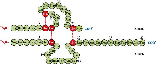 Figure: 1.10. Disulfide bonds in the structure of insulin
5. Supersecondary structure of proteins.In proteins that differ in their primary structure and functions, sometimes similar combinations and arrangement of secondary structures,which are called supersecondary structure. It occupies an intermediate position between the secondary and tertiary structures, since this is a specific combination of elements of the secondary structure in the formation of the tertiary structure of the protein. Supersecondary structures have specific names such as “α-helix-turn-a-helix”, “leucine zipper”, “zinc fingers”, etc. Such supersecondary structures are characteristic of DNA-binding proteins.
Leucine zipper.This kind of supersecondary structure is used to connect two proteins. On the surface of interacting proteins, there are α-helical regions containing at least four leucine residues. Leucine residues in the α-helix are located six amino acids apart from each other. Since each turn of the α-helix contains 3.6 amino acid residues, leucine radicals are located on the surface of every second turn. Leucine residues of the α-helix of one protein can interact with leucine residues of another protein (hydrophobic interactions), linking them together (Fig. 1.11.). Many DNA-binding proteins function as part of oligomeric complexes, where individual subunits bind to each other with "leucine fasteners".
 Figure: 1.11. "Leucine zipper" between the α-helical regions of two proteins
An example of such proteins is histones. Histones- nuclear proteins, which include a large number of positively charged amino acids - arginine and lysine (up to 80%). Histone molecules are combined into oligomeric complexes containing eight monomers with the help of "leucine fasteners", despite the significant charge of the same name of these molecules.
"Zinc finger"- a variant of the supersecondary structure, characteristic of DNA-binding proteins, has the form of an elongated fragment on the protein surface and contains about 20 amino acid residues (Fig. 1.12). The shape of the "elongated finger" is supported by a zinc atom linked to the radicals of four amino acids - two cysteine \u200b\u200bresidues and two histidine residues. In some cases, instead of histidine residues, there are cysteine \u200b\u200bresidues. Two closely spaced cysteine \u200b\u200bresidues are separated from the other two Gisili residues by a sequence of about 12 amino acid residues. This region of the protein forms an α-helix, the radicals of which can specifically bind to the regulatory regions of the major groove of DNA. The binding specificity of an individual
 Figure: 1.12. The primary structure of a region of DNA-binding proteins that form the structure of the "zinc finger" (letters indicate the amino acids that make up this structure)
regulatory DNA-binding protein depends on the sequence of amino acid residues located in the "zinc finger". Such structures contain, in particular, receptors for steroid hormones involved in the regulation of transcription (reading information from DNA to RNA).
TOPIC 1.2. BASES OF FUNCTIONING OF PROTEINS. MEDICINES AS LIGANDS AFFECTING THE FUNCTION OF PROTEINS
1. The active center of the protein and its interaction with the ligand.During the formation of a tertiary structure on the surface of a functionally active protein, usually in a depression, a region is formed that is formed by amino acid radicals that are far from each other in the primary structure. This site, which has a unique structure for a given protein and is able to specifically interact with a certain molecule or a group of similar molecules, is called the protein-ligand binding site or active site. Ligands are molecules that interact with proteins.
High specificitythe interaction of the protein with the ligand is provided by the complementarity of the structure of the active center to the structure of the ligand.
Complementarityis the spatial and chemical correspondence of interacting surfaces. The active center should not only spatially correspond to the ligand included in it, but also bonds (ionic, hydrogen, and also hydrophobic interactions) should form between the functional groups of the radicals included in the active center and the ligand, which hold the ligand in the active center (Fig.1.13 ).
 Figure: 1.13. Complementary interaction of a protein with a ligand
Some ligands, by attaching to the active center of the protein, play an auxiliary role in the functioning of proteins. Such ligands are called cofactors, and proteins containing a non-protein part are called complex proteins(as opposed to simple proteins, consisting only of the protein part). The non-protein part, firmly attached to the protein, is called prosthetic group.For example, the composition of myoglobin, hemoglobin and cytochromes contains a prosthetic group firmly attached to the active center - heme, which contains an iron ion. The complex proteins that contain heme are called hemoproteins.
When specific ligands are attached to proteins, the function of these proteins is manifested. So, albumin - the most important protein in blood plasma - manifests its transport function by attaching hydrophobic ligands such as fatty acids, bilirubin, some drugs, etc. to the active center (Fig.1.14)
Ligands interacting with the three-dimensional structure of the peptide chain can be not only low molecular weight organic and inorganic molecules, but also macromolecules:
DNA (examples of DNA-binding proteins discussed above);
Polysaccharides;
 Figure: 1.14. Relationship between genotype and phenotype
The unique primary structure of human proteins, encoded in the DNA molecule, is realized in cells in the form of a unique conformation, structure of the active center and functions of proteins
In these cases, the protein recognizes a certain ligand site commensurate with and complementary to the binding site. So on the surface of hepatocytes there are receptor proteins for the hormone insulin, which also has a protein structure. The interaction of insulin with the receptor causes a change in its conformation and activation of signaling systems, leading to the accumulation of nutrients in hepatocytes after eating.
Thus, the functioning of proteins is based on the specific interaction of the active center of the protein with the ligand.
2. Domain structure and its role in the functioning of proteins.Long polypeptide chains of globular proteins often fold into several compact, relatively independent regions. They have an independent tertiary structure, similar to that of globular proteins, and are called domains.Due to the domain structure of proteins, their tertiary structure is more easily formed.
In domain proteins, ligand binding sites are often located between domains. Thus, trypsin is a proteolytic enzyme that is produced by the exocrine part of the pancreas and is necessary for the digestion of food proteins. It has a two-domain structure, and the binding site of trypsin with its ligand, a food protein, is located in the groove between the two domains. In the active center, conditions are created that are necessary for the effective binding of a specific site of the food protein and the hydrolysis of its peptide bonds.
When the active center interacts with the ligand, different domains in a protein can move relative to each other (Fig. 1.15).
Hexokinase- an enzyme that catalyzes the phosphorylation of glucose by means of ATP. The active site of the enzyme is located in the cleft between the two domains. When hexokinase binds to glucose, the surrounding domains close and the substrate is trapped, where phosphorylation occurs (see Fig. 1.15).
 Figure: 1.15. Binding of Hexokinase Domains to Glucose
In some proteins, domains perform independent functions by binding to various ligands. These proteins are called multifunctional.
3. Drugs - ligands that affect the function of proteins.The interaction of proteins with ligands is specific. However, due to the conformational lability of the protein and its active center, it is possible to select another substance that could also interact with the protein in the active center or other part of the molecule.
A substance similar in structure to a natural ligand is called structural analogue of the ligandor an unnatural ligand. It also interacts with a protein in the active site. The structural analogue of the ligand can both enhance the function of the protein (agonist),so reduce it (antagonist).The ligand and its structural analogs compete with each other for binding to the protein at the same center. Such substances are called competitive modulators(regulators) of protein functions. Many drugs act as protein inhibitors. Some of them are obtained by chemical modification of natural ligands. Protein inhibitors can be drugs and poisons.
Atropine is a competitive inhibitor of M-cholinergic receptors.Acetylcholine is a neurotransmitter for the transmission of nerve impulses through cholinergic synapses. To conduct excitation, acetylcholine released into the synaptic cleft must interact with a protein - the receptor of the postsynaptic membrane. Two types found cholinergic receptors:
M-receptor,in addition to acetylcholine, it selectively interacts with muscarin (fly agaric toxin). M - cholinergic receptors are present on smooth muscles and, when interacting with acetylcholine, cause their contraction;
H-receptor,specifically binding to nicotine. H-cholinergic receptors are found in the synapses of striated skeletal muscles.
Specific inhibitor M-cholinergic receptorsis atropine. It is found in belladonna and henbane plants.
 Atropine has in the structure functional groups similar to acetylcholine and their spatial arrangement, therefore, it belongs to competitive inhibitors of M-cholinergic receptors. Considering that the binding of acetylcholine to M-cholinergic receptors causes contraction of smooth muscles, atropine is used as a medicine that relieves muscle spasms. (antispasmodic).So, the use of atropine is known to relax the eye muscles when looking at the fundus, as well as to relieve spasms in gastrointestinal colic. M-cholinergic receptors are also present in the central nervous system (CNS), therefore, large doses of atropine can cause an unwanted reaction from the central nervous system: motor and mental agitation, hallucinations, convulsions.
Ditilin is a competitive agonist of H-cholinergic receptors that inhibits the function of neuromuscular synapses.
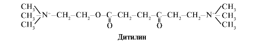 Skeletal muscle neuromuscular synapses contain H-cholinergic receptors. Their interaction with acetylcholine leads to muscle contractions. Some surgical procedures, as well as endoscopic examinations, use drugs that cause relaxation of skeletal muscles (muscle relaxants).These include dityline, which is a structural analog of acetylcholine. It attaches to H-cholinergic receptors, but unlike acetylcholine, it is very slowly destroyed by the enzyme, acetylcholinesterase. As a result of prolonged opening of ion channels and persistent depolarization of the membrane, the conduction of the nerve impulse is disrupted and muscle relaxation occurs. Initially, these properties were found in curare poison, therefore such drugs are called curariform.
TOPIC 1.3. DENATURATION OF PROTEINS AND THE POSSIBILITY OF THEIR SPONTANEOUS RENATIVATION
1. Since the native conformation of proteins is maintained due to weak interactions, changes in the composition and properties of the environment surrounding the protein, exposure to chemical reagents and physical factors cause a change in their conformation (property of conformational lability). The breaking of a large number of bonds leads to the destruction of the native conformation and denaturation of proteins.
Protein denaturationis the destruction of their native conformation under the action of denaturing agents, caused by the breaking of weak bonds that stabilize the spatial structure of the protein. Denaturation is accompanied by the destruction of the unique three-dimensional structure and active center of the protein and the loss of its biological activity (Fig. 1.16).
All denatured molecules of one protein acquire a random conformation that differs from other molecules of the same protein. The amino acid radicals that form the active center are spatially distant from each other, i.e. the specific binding site of the protein with the ligand is destroyed. During denaturation, the primary structure of proteins remains unchanged.
The use of denaturing agents in biological research and medicine.In biochemical studies, before the determination of low molecular weight compounds in biological material, proteins are usually removed from the solution first. For this purpose, trichloroacetic acid (TCA) is most often used. After adding TCA to the solution, denatured proteins precipitate and are easily removed by filtration (Table 1.1.)
In medicine, denaturing agents are often used to sterilize medical instruments and materials in autoclaves (denaturing agent - high temperature) and as antiseptics (alcohol, phenol, chloramine) to treat contaminated surfaces containing pathogenic microflora.
2. Spontaneous protein renewal- proof of the determinism of the primary structure, conformation and function of proteins. Individual proteins are products of one gene that have an identical amino acid sequence and acquire the same conformation in the cell. The fundamental conclusion that the primary structure of a protein already contains information about its conformation and function was made on the basis of the ability of some proteins (in particular, ribonuclease and myoglobin) to spontaneously regenerate - to restore their native conformation after denaturation.
The formation of spatial structures of a protein is carried out by the method of self-assembly - a spontaneous process in which a polypeptide chain, which has a unique primary structure, tends to accept a conformation with the lowest free energy in solution. The ability to renew proteins that retain their primary structure after denaturation was described in an experiment with the enzyme ribonuclease.
Ribonuclease is an enzyme that breaks down the bonds between individual nucleotides in an RNA molecule. This globular protein has a single polypeptide chain, the tertiary structure of which is stabilized by many weak and four disulfide bonds.
Treatment of ribonuclease with urea, which destroys hydrogen bonds in the molecule, and a reducing agent that breaks disulfide bonds, leads to denaturation of the enzyme and the loss of its activity.
Removal of denaturing agents by dialysis results in restoration of protein conformation and function, i.e. to the renewal. (fig. 1.17).
 Figure: 1.17. Denaturation and renaissance of ribonuclease
A - native conformation of ribonuclease, in the tertiary structure of which there are four disulfide bonds; B - denatured ribonuclease molecule;
B - revived ribonuclease molecule with reduced structure and function
1. Complete table 1.2.
Table 1.2. Classification of amino acids by radical polarity
2. Write the formula for tetrapeptide:
Asp - Pro - Fen - Liz
a) isolate in the peptide the repeating groups that form the peptide backbone and variable groups represented by amino acid radicals;
b) designate the N- and C-ends;
c) underline peptide bonds;
d) write another peptide consisting of the same amino acids;
e) count the number of possible variants of a tetrapeptide with a similar amino acid composition.
3. Explain the role of the primary structure of proteins by the example of a comparative analysis of two structurally similar and evolutionarily close peptide hormones of the mammalian neurohypophysis - oxytocin and vasopressin (Table 1.3).
Table 1.3. Structure and function of oxytocin and vasopressin
 For this:
a) compare the composition and amino acid sequence of the two peptides;
b) find the similarity of the primary structure of two peptides and the similarity of their biological action;
c) find the differences in the structure of the two peptides and the difference in their functions;
d) draw a conclusion about the influence of the primary structure of peptides on their functions.
4. Describe the main stages in the formation of the conformation of globular proteins (secondary, tertiary structures, the concept of a supersecondary structure). Indicate the types of bonds involved in the formation of protein structures. Which amino acid radicals can participate in the formation of hydrophobic interactions, ionic, hydrogen bonds.
Give examples.
5. Give a definition of the concept of "conformational lability of proteins", indicate the reasons for its existence and significance.
6. Expand the meaning of the following phrase: "The functioning of proteins is based on their specific interaction with the ligand", using the terms and explaining their meaning: protein conformation, active center, ligand, complementarity, protein function.
7. Use one example to explain what domains are and what their role in proteins is.
TASKS FOR SELF-CONTROL
1. Establish correspondence.
Functional group in the amino acid radical:
A. Carboxyl group B. Hydroxyl group C Guanidine group D. Thiol group D. Amino group
2. Choose the correct answers.
Amino acids with polar uncharged radicals are:
A. Cis B. Asn
B. Glu G. Three
3. Choose the correct answers.
Amino acid radicals:
A. Provide specificity of the primary structure B. Participate in the formation of the tertiary structure
B. Located on the surface of the protein, affect its solubility D. Form an active center
E. Participate in the formation of peptide bonds
4. Choose the correct answers.
Hydrophobic interactions can form between amino acid radicals:
A. Tre Lei B. Pro Three
B. Met Ile G. Tir Ala D. Val Fen
5. Choose the correct answers.
Ionic bonds can form between amino acid radicals:
A. Gln Asp B. Apr Liz
B. Liz Glu G. Gies Asp D. Asn Apr
6. Choose the correct answers.
Hydrogen bonds can form between amino acid radicals:
A. Ser Gln B. Cis Tre
B. Asp Liz G. Glu Asp D. Asn Tre
7. Establish correspondence.
The type of bond involved in the formation of the protein structure:
A. Primary structure B. Secondary structure
B. Tertiary structure
D. Supersecondary structure E. Conformation.
1. Hydrogen bonds between the atoms of the peptide backbone
2. Weak bonds between functional groups of amino acid radicals
3. Relationships between α-amino and α-carboxyl groups of amino acids
8. Choose the correct answers. Trypsin:
A. Proteolytic enzyme B. Contains two domains
B. Hydrolyzes starch
D. The active center is located between the domains. D. Consists of two polypeptide chains.
9. Choose the correct answers. Atropine:
A. Neurotransmitter
B. Structural analogue of acetylcholine
B. Interacts with H-cholinergic receptors
D. Strengthens the conduction of nerve impulses through cholinergic synapses
D. Competitive inhibitor of M-cholinergic receptors
10. Choose the correct statements. In proteins:
A. Primary structure contains information about the structure of its active center
B. The active center is formed at the level of the primary structure
B. The conformation is rigidly fixed by covalent bonds
D. The active site can interact with a group of similar ligands
due to the conformational lability of proteins D. Changes in the environment can affect the affinity of the active
center to ligand
1.1-C, 2-D, 3-B.
3.A, B, C, D.
7.1-B, 2-D, 3-A.
8.A, B, C, D.
BASIC TERMS AND CONCEPTS
1. Protein, polypeptide, amino acids
2. Primary, secondary, tertiary protein structure
3. Conformation, native protein conformation
4. Covalent and weak bonds in a protein
5. Conformational lability
6. Active center of protein
7. Ligands
8. Protein folding
9. Structural analogs of ligands
10. Domain proteins
11. Simple and complex proteins
12. Protein denaturation, denaturing agents
13. Renovation of proteins
Solve tasks
"Structural organization of proteins and the basis of their functioning"
1. The main function of the protein, hemoglobin A (HbA), is to transport oxygen to tissues. In the human population, multiple forms of this protein with altered properties and functions are known - the so-called abnormal hemoglobins. For example, it was found that hemoglobin S, found in the erythrocytes of sickle cell anemia (HbS) patients, has low solubility under conditions of low oxygen partial pressure (as is the case in venous blood). This leads to the formation of aggregates of this protein. The protein loses its function, precipitates, and red blood cells acquire an irregular shape (some of them form a sickle) and are destroyed faster than usual in the spleen. As a result, sickle cell anemia develops.
The only difference in the primary structure of HbA was found in the N-terminal region of the β-chain of hemoglobin. Compare the N-terminus of the β-chain and show how changes in the primary structure of a protein affect its properties and functions.
 For this:
a) write the amino acid formulas by which HBA differ and compare the properties of these amino acids (polarity, charge).
b) draw a conclusion about the reason for the decrease in solubility and impaired oxygen transport in the tissue.
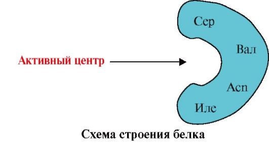
2. The figure shows a schematic diagram of the structure of a protein that has a ligand-binding site (active site). Explain why a protein is selective in its choice of ligand. For this:
a) remember what the active center of a protein is, and consider the structure of the active center of the protein shown in the figure;
b) write the formulas of amino acid radicals that make up the active center;
c) draw a ligand that could specifically interact with the active center of the protein. Indicate on it the functional groups capable of forming bonds with the amino acid radicals that make up the active center;
d) indicate the types of bonds that arise between the ligand and the amino acid radicals of the active center;
e) explain what the specificity of the interaction of the protein with the ligand is based on.
 3.
The figure shows the active site of the protein and several ligands.
3.
The figure shows the active site of the protein and several ligands.
Determine which ligand is most likely to interact with the active site of the protein and why.
 What types of bonds arise during the formation of a protein-ligand complex?
4. Structural analogs of natural protein ligands can be used as drugs to alter the activity of proteins.
Acetylcholine is a transmitter of excitation in neuromuscular synapses. When acetylcholine interacts with proteins - receptors of the postsynaptic membrane of skeletal muscles, ion channels open and muscle contraction. Ditilin is a medicine used in some operations to relax muscles, since it disrupts the transmission of nerve impulses through the neuromuscular synapses. Explain the mechanism of action of ditilin as a muscle relaxant drug. For this:
a) write the formulas of acetylcholine and ditilin and compare their structures;
b) describe the mechanism of the relaxing action of ditilin.
5. In some diseases, the patient's body temperature rises, which is considered as a protective reaction of the body. However, high temperatures are detrimental to body proteins. Explain why at temperatures above 40 ° C, the function of proteins is disrupted and a threat to human life arises. To do this, remember:
1) The structure of proteins and bonds that hold its structure in the native conformation;
2) How does the structure and function of proteins change with increasing temperature?;
3) What is homeostasis and why is it important for maintaining human health.
Modular unit 2 OLIGOMERIC PROTEINS AS TARGETS OF REGULATORY IMPACT. STRUCTURAL AND FUNCTIONAL PROTEIN VARIETY. PROTEIN SEPARATION AND PURIFICATION METHODS
Learning Objectives To be able to:
1. Use knowledge about the features of the structure and functions of oligomeric proteins to understand the adaptive mechanisms of regulation of their functions.
2. Explain the role of chaperones in the synthesis and maintenance of protein conformation under cell conditions.
3. Explain the variety of manifestations of life by the variety of structures and functions of proteins synthesized in the body.
4. To analyze the relationship between the structure of proteins and their function by examples of comparing related hemoproteins - myoglobin and hemoglobin, as well as representatives of five classes of proteins of the immunoglobulin family.
5. Apply knowledge about the peculiarities of the physicochemical properties of proteins to select methods for their purification from other proteins and impurities.
6. Interpret the results of the quantitative and qualitative composition of blood plasma proteins to confirm or clarify the clinical diagnosis.
Know:
1. Features of the structure of oligomeric proteins and adaptive mechanisms of regulation of their functions on the example of hemoglobin.
2. The structure and function of chaperones and their importance for maintaining the native conformation of proteins under cell conditions.
3. Principles of combining proteins into families according to the similarity of their conformation and functions on the example of immunoglobulins.
4. Methods for the separation of proteins based on the peculiarities of their physicochemical properties.
5. Electrophoresis of blood plasma as a method for assessing the qualitative and quantitative composition of proteins.
TOPIC 1.4. FEATURES OF THE STRUCTURE AND FUNCTIONING OF OLIGOMERIC PROTEINS ON THE EXAMPLE OF HEMOGLOBIN
1. Many proteins contain several polypeptide chains. Such proteins are called oligomeric,and individual chains - protomers.Protomers in oligomeric proteins are linked by many weak non-covalent bonds (hydrophobic, ionic, hydrogen). Interaction
protomers is carried out thanks complementaritytheir contact surfaces.
The number of protomers in oligomeric proteins can vary greatly: hemoglobin contains 4 protomers, the aspartate aminotransferase enzyme contains 12 protomers, and the tobacco mosaic virus protein contains 2,120 protomers linked by non-covalent bonds. Consequently, oligomeric proteins can have very high molecular weights.
The interaction of one protomer with others can be considered as a special case of the interaction of a protein with a ligand, since each protomer serves as a ligand for other protomers. The number and method of combining protomers in a protein is called quaternary protein structure.
Proteins can include protomers of the same or different structure, for example, homodimers are proteins containing two identical protomers, and heterodimers are proteins containing two different protomers.
If proteins contain different protomers, then different structures of binding sites with different ligands can be formed on them. When the ligand binds to the active site, the function of this protein is manifested. The center located on another protomer is called allosteric (other than the active one). By contacting an allosteric ligand or effector,it performs a regulatory function (Fig. 1.18). The interaction of the allosteric center with the effector causes conformational changes in the structure of the entire oligomeric protein due to its conformational lability. This affects the affinity of the active site for a specific ligand and regulates the function of this protein. The change in the conformation and function of all protomers upon interaction of an oligomeric protein with at least one ligand is called cooperative conformational changes. Effectors that enhance protein function are called activators,and the effectors that inhibit its function - inhibitors.
Thus, oligomeric proteins, as well as proteins with a domain structure, have a new property in comparison with monomeric proteins - the ability to allosteric regulation of functions (regulation by the attachment of various ligands to the protein). This can be seen by comparing the structures and functions of two closely related complex proteins, myoglobin and hemoglobin.
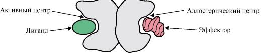 Figure: 1.18. Dimeric protein structure diagram
2. The formation of spatial structures and the functioning of myoglobin.
Myoglobin (MB) is a protein found in red muscles, the main function of which is to create O 2 reserves, which are necessary for intensive muscular work. MB is a complex protein containing a protein part - apoMv and a non-protein part - heme. The primary structure of apoMv determines its compact globular conformation and the structure of the active site, to which the non-protein part of myoglobin, heme, is attached. Oxygen coming from the blood to the muscles binds to Fe + 2 heme in myoglobin. MB is a monomeric protein with a very high affinity for O 2, therefore, the release of oxygen by myoglobin occurs only during intense muscular work, when the partial pressure of O 2 drops sharply.
Formation of the MB conformation.In red muscles on ribosomes, during translation, the primary structure of MB is synthesized, represented by a specific sequence of 153 amino acid residues. The secondary structure of MB contains eight α-helices, called the Latin letters from A to H, between which there are non-helical sections. The tertiary structure of MB has the form of a compact globule, in the deepening of which between the F and E α-helices there is an active center (Fig. 1.19).
 Figure: 1.19. Myoglobin structure
3. Features of the structure and functioning of the active center Мв.The active center of MB is formed mainly by hydrophobic amino acid radicals that are far from each other in the primary structure (for example, Three 3 9 and Phen 138) Poorly water-soluble ligands - heme and O 2 - attach to the active center. Heme is a specific ligand of apoMv (Fig. 1.20), which is based on four pyrrole rings connected by metenyl bridges; the Fe + 2 atom is located in the center, connected to the nitrogen atoms of the pyrrole rings by four coordination bonds. In the active center of MB, in addition to hydrophobic amino acid radicals, there are also residues of two amino acids with hydrophilic radicals - Gis E 7(Gis 64) and Gis F 8(Gis 93) (Fig. 1.21).
 Figure: 1.20. The structure of heme - the non-protein part of myoglobin and hemoglobin
 Figure: 1.21. Location of heme and O 2 in the active center of apomyoglobin and hemoglobin protomers
Heme through an iron atom is covalently bound to His F 8. O 2 attaches to the gland on the other side of the heme plane. His E 7 is necessary for the correct orientation of O 2 and facilitates the addition of oxygen to Fe + 2 heme
Gis F 8forms a coordination bond with Fe + 2 and firmly fixes the heme in the active center. Gis E 7is necessary for the correct orientation in the active center of another ligand, O 2, when it interacts with Fe + 2 heme. The microenvironment of heme creates conditions for strong but reversible binding of O 2 with Fe +2 and prevents water from entering the hydrophobic active center, which can lead to its oxidation into Fe + 3.
The monomeric structure of MB and its active center determines the high affinity of the protein for O 2.
4. Oligomeric structure of HB and regulation of the affinity of HB to O 2 ligands. Human hemoglobins- a family of proteins, as well as myoglobin, related to complex proteins (hemoproteins). They have a tetrameric structure and contain two α-chains, but differ in the structure of the other two polypeptide chains (2α-, 2x-chains). The structure of the second polypeptide chain determines the features of the functioning of these forms of HB. About 98% of the hemoglobin of erythrocytes of an adult is hemoglobin A(2α-, 2p-chains).
During intrauterine development, two main types of hemoglobins function: embryonic HB(2α, 2ε), which is found in the early stages of fetal development, and hemoglobin F (fetal)- (2α, 2γ), which replaces early fetal hemoglobin in the sixth month of intrauterine development and only after birth is replaced by HB A.
HB A is a protein related to myoglobin (MB) and is found in the erythrocytes of an adult. The structure of its individual protomers is similar to that of myoglobin. The secondary and tertiary structures of myoglobin and hemoglobin protomers are very similar, despite the fact that only 24 amino acid residues are identical in the primary structure of their polypeptide chains (the secondary structure of hemoglobin protomers, like myoglobin, contains eight α-helices, denoted by Latin letters from A to H , and the tertiary structure looks like a compact globule). But unlike myoglobin, hemoglobin has an oligomeric structure, consists of four polypeptide chains connected by non-covalent bonds (Figure 1.22).
Each HB protomer is associated with a non-protein part — heme and adjacent protomers. The connection of the protein part of HB with heme is similar to that of myoglobin: in the active center of the protein, the hydrophobic parts of the heme are surrounded by hydrophobic amino acid radicals, with the exception of His F 8 and His E 7, which are located on both sides of the heme plane and play a similar role in the functioning of the protein and its binding with oxygen (see the structure of myoglobin).
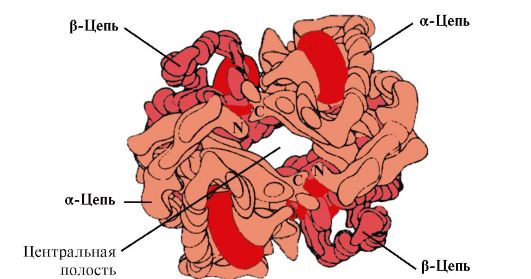 Figure: 1.22. Oligomeric structure of hemoglobin
Besides, Gis E 7performs an important additional rolein the functioning of NV. Free heme has a 25,000 times higher affinity for CO than for O 2. CO is formed in small amounts in the body and, given its high affinity for heme, it could disrupt the transport of O 2 necessary for the life of cells. However, in the composition of hemoglobin, the affinity of heme for carbon monoxide exceeds the affinity for O 2 by only 200 times due to the presence of His E 7 in the active center. The remainder of this amino acid creates optimal conditions for the binding of heme with O 2 and weakens the interaction of heme with CO.
5. The main function of HB is the transport of O 2 from the lungs to the tissue.In contrast to monomeric myoglobin, which has a very high affinity for O 2 and performs the function of storing oxygen in red muscles, the oligomeric structure of hemoglobin provides:
1) rapid saturation of Hb with oxygen in the lungs;
2) the ability of HB to donate oxygen in tissues at a relatively high partial pressure of O 2 (20-40 mm Hg);
3) the ability to regulate the affinity of HB to O 2.
6. Cooperative changes in the conformation of hemoglobin protomers accelerate the binding of O 2 in the lungs and its release to the tissues. In the lungs, the high partial pressure of O 2 promotes its binding to HB in the active center of four protomers (2α and 2β). The active center of each protomer, as in myoglobin, is located between two α-helices (F and E) in a hydrophobic pocket. It contains a non-protein part - heme, attached to the protein part by many weak hydrophobic interactions and one strong bond between Fe 2 + heme and His F 8 (see Fig. 1.21).
In deoxyhemoglobin, due to this bond with His F 8, the Fe 2 + atom protrudes from the heme plane towards histidine. The binding of O 2 with Fe 2 + occurs on the other side of the heme in the region of His E 7 with the help of a single free coordination bond. His E 7 provides optimal conditions for the binding of O 2 to heme iron.
The attachment of O 2 to the Fe +2 atom of one protomer causes it to move into the heme plane, followed by the histidine residue bound to it.
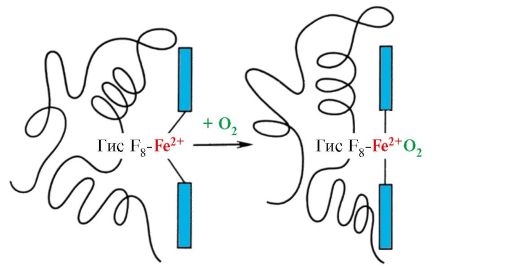 Figure: 1.23. Change in the conformation of the hemoglobin protomer when combined with O 2
This leads to a change in the conformation of all polypeptide chains due to their conformational lability. Changing the conformation of other chains facilitates their interaction with the following O 2 molecules.
The fourth O 2 molecule binds to hemoglobin 300 times more easily than the first (Fig. 1.24).
 Figure: 1.24. Cooperative changes in the conformation of hemoglobin protomers upon its interaction with О 2
In tissues, each subsequent O 2 molecule is cleaved off more easily than the previous one, also due to cooperative changes in the conformation of protomers.
7. CO 2 and H +, formed during the catabolism of organic substances, reduce the affinity of hemoglobin to O 2 in proportion to their concentration. The energy required for the functioning of cells is produced mainly in the mitochondria during the oxidation of organic substances using O 2 delivered from the lungs by hemoglobin. As a result of the oxidation of organic substances, the final products of their decay are formed: CO 2 and K 2 O, the amount of which is proportional to the intensity of the ongoing oxidation processes.
CO 2 diffusion from cells into the blood and penetrates into erythrocytes, where it is converted into carbonic acid under the action of the enzyme carbanhydrase. This weak acid dissociates into a proton and bicarbonate ion.
H + are able to join radicals of His 14 6 in the α- and β-chains of hemoglobin, i.e. in areas remote from the heme. Protonation of hemoglobin reduces its affinity for O 2, promotes the elimination of O 2 from oxyHb, the formation of deoxyHb, and increases the supply of oxygen to tissues in proportion to the amount of protons formed (Fig. 1.25).
An increase in the amount of liberated oxygen, depending on an increase in the concentration of H + in erythrocytes, is called the Bohr effect (after the Danish physiologist Christian Bohr, who first discovered this effect).
In the lungs, the high partial pressure of oxygen promotes its binding to deoxyHB, which reduces the protein's affinity for H +. The released protons under the action of carbonic anhydrase interact with bicarbonates with the formation of CO 2 and H 2 O

 Figure: 1.25. Dependence of the affinity of Нв for О 2 on the concentration of СО 2 and protons (Bohr effect):
AND- the influence of the concentration of CO 2 and H + on the release of O 2 from the complex with HB (Bohr effect); B- oxygenation of deoxyhemoglobin in the lungs, the formation and release of CO 2.
The resulting CO 2 enters the alveolar space and is removed with exhaled air. Thus, the amount of oxygen released by hemoglobin in tissues is regulated by the products of catabolism of organic substances: the more intensive the decomposition of substances, for example, during physical exertion, the higher the concentration of CO 2 and H + and the more oxygen the tissues receive as a result of a decrease in the affinity of Hb to O 2.
8. Allosteric regulation of the Hb affinity for O 2 by the ligand - 2,3-bis-phosphoglycerate.In erythrocytes, an allosteric ligand of hemoglobin, 2,3-bisphosphoglycerate (2,3-BFG), is synthesized from the glucose oxidation product, 1,3-bisphosphoglycerate. Under normal conditions, the concentration of 2,3-BFG is high and comparable to the concentration of HB. 2,3-BFG has a strong negative charge of -5.
 Bisphosphoglycerate in the capillaries of tissues, binding with deoxyhemoglobin, increases the release of oxygen into the tissue, reducing the affinity of HB for O 2.
In the center of the tetrameric hemoglobin molecule there is a cavity. It is formed by amino acid residues of all four protomers (see Fig. 1.22). In tissue capillaries, protonation of HB (Bohr effect) leads to a breakdown of the bond between heme iron and O 2. In a molecule
deoxyhemoglobin, in comparison with oxyhemoglobin, additional ionic bonds arise that connect protomers, as a result of which the dimensions of the central cavity increase in comparison with oxyhemoglobin. The central cavity is the place where 2,3-BFG attaches to hemoglobin. Due to the difference in the size of the central cavity, 2,3-BFG can only attach to deoxyhemoglobin.
2,3-BFG interacts with hemoglobin in a region remote from the active centers of the protein and belongs to allosteric(regulatory) ligands, and the central cavity of HB is allosteric center.2,3-BFG has a strong negative charge and interacts with five positively charged groups of two β-chains of HB: the N-terminal α-amino group Val and the radicals Lys 82 His 143 (Fig. 1.26).
 Figure: 1.26. BPH in the central cavity of deoxyhemoglobin
BPH binds to three positively charged groups in each β-chain.
In tissue capillaries, the resulting deoxyhemoglobin interacts with 2,3-BFG and ionic bonds are formed between the positively charged radicals of the β-chains and the negatively charged ligand, which change the conformation of the protein and reduce the affinity of HB for O 2. A decrease in the affinity of HB for O 2 contributes to a more efficient release of O 2 into the tissue.
In the lungs at high partial pressures, oxygen interacts with HB, attaching itself to the heme iron; at the same time, the conformation of the protein changes, the central cavity decreases and 2,3-BPG is displaced from the allosteric center
Thus, oligomeric proteins have properties that are new compared to monomeric proteins. Attachment of ligands at sites
spatially distant from each other (allosteric), capable of causing conformational changes in the entire protein molecule. Due to the interaction with regulatory ligands, the conformation changes and the function of the protein molecule adapts to changes in the environment.
TOPIC 1.5. MAINTENANCE OF THE NATIVE CONFORMATION OF PROTEINS UNDER CELL CONDITIONS
In cells, during the synthesis of polypeptide chains, their transport across membranes to the corresponding sections of the cell, in the process of folding (the formation of a native conformation) and during the assembly of oligomeric proteins, as well as during their functioning, intermediate, prone to aggregation, unstable conformations appear in the structure of proteins. Hydrophobic radicals, in the native conformation usually hidden inside the protein molecule, find themselves on the surface in an unstable conformation and tend to combine with the same poorly water-soluble groups of other proteins. In the cells of all known organisms, special proteins have been found that ensure optimal folding of cell proteins, stabilize their native conformation during functioning and, which is especially important, maintain the structure and function of intracellular proteins in case of homeostasis disturbance. These proteins are named "Chaperones",which is translated from French means "nanny".
1. Molecular chaperones and their role in preventing protein denaturation.
Chaperones (III) are classified by subunit mass. High molecular weight chaperones have a mass of 60 to 110 kDa. Among them, the most studied are three classes: Sh-60, Sh-70 and Sh-90. Each class includes a family of related proteins. Thus, Sh-70 contains proteins with molecular weights from 66 to 78 kDa. Low molecular weight chaperones have a molecular weight of 40 to 15 kDa.
Among the chaperones are distinguished constitutiveproteins, high basal synthesis of which does not depend on stress effects on the cells of the body, and inducible,the synthesis of which under normal conditions is weak, but increases sharply under stress. Inducible chaperones are also referred to as "heat shock proteins" because they were first discovered in cells exposed to high temperatures. In cells, due to the high concentration of proteins, spontaneous renewal of partially denatured proteins is difficult. III-70 can prevent the incipient process of denaturation and help restore the native conformation of proteins. Molecular chaperones-70- a highly conserved class of proteins found in all parts of the cell: cytoplasm, nucleus, endoplasmic reticulum, mitochondria. At the carboxyl end of the only III-70 polypeptide chain, there is a region that is a groove that can interact with peptides of length
from 7 to 9 amino acid residues enriched in hydrophobic radicals. Such regions in globular proteins occur approximately every 16 amino acids. III-70 are able to protect proteins from thermal inactivation and restore the conformation and activity of partially denatured proteins.
2. The role of chaperones in protein folding.When proteins are synthesized on the ribosome, the N-terminal region of the polypeptide is synthesized earlier than the C-terminal region. The complete amino acid sequence of the protein is required to form the native conformation. In the process of protein synthesis, chaperones-70, due to the structure of their active center, are able to close the aggregation-prone regions of the polypeptide enriched in hydrophobic amino acid radicals until the synthesis is complete (Figure 1.27, A).
 Figure: 1.27. Participation of chaperones in protein folding
A - participation of chaperones-70 in the prevention of hydrophobic interactions between regions of the synthesized polypeptide; B - formation of native protein conformation in the chaperone complex
Many high-molecular-weight proteins with a complex conformation, for example, a domain structure, fold in a special space formed by III-60. Sh-60they function as an oligomeric complex consisting of 14 subunits. They form two hollow rings, each of which consists of seven subunits, these rings are connected to each other. Each III-60 subunit consists of three domains: apical (apical), enriched in hydrophobic radicals facing the ring cavity, intermediate and equatorial (Fig. 1.28).
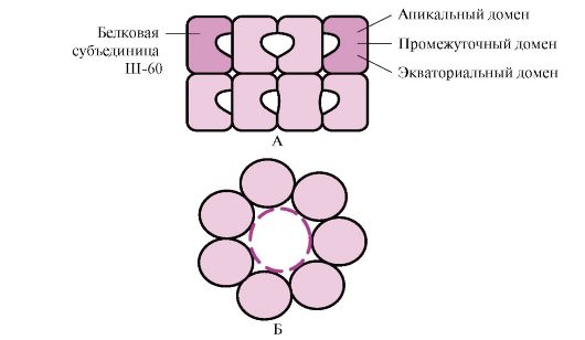 Figure: 1.28. The structure of the chaperonin complex, consisting of 14 Sh-60
A - side view; B - top view
The synthesized proteins, which have on the surface elements characteristic of unfolded molecules, in particular hydrophobic radicals, enter the cavity of the chaperone rings. In the specific environment of these cavities, an enumeration of possible conformations occurs until the only one, energetically most favorable, is found (Fig. 1.27, B). The formation of conformations and the release of protein is accompanied by hydrolysis of ATP in the equatorial region. Typically, such chaperone-dependent folding requires a significant amount of energy.
In addition to participating in the formation of the three-dimensional structure of proteins and the revival of partially denatured proteins, chaperones are also required for such fundamental processes as the assembly of oligomeric proteins, recognition and transport of denatured proteins into lysosomes, transport of proteins across membranes, and participation in the regulation of the activity of protein complexes.
TOPIC 1.6. VARIETY OF PROTEINS. PROTEIN FAMILIES BY THE EXAMPLE OF IMMUNOGLOBULINS
1. Proteins play a decisive role in the life of individual cells and the entire multicellular organism, and their functions are surprisingly diverse. This is determined by the peculiarities of the primary structure and conformations of proteins, the uniqueness of the structure of the active center, and the ability to bind specific ligands.
Only a very small fraction of all possible variants of peptide chains can assume a stable spatial structure; most
of them can take many conformations with approximately the same Gibbs energy, but with different properties. The primary structure of most of the known proteins selected by biological evolution provides exceptional stability of one of the conformations that determines the features of the functioning of this protein.
2. Protein families.Within the same biological species, substitutions of amino acid residues can lead to the appearance of different proteins that perform related functions and have homologous amino acid sequences. Such related proteins have strikingly similar conformations: the number and interposition of α-helices and / or β-structures, most of the turns and bends of the polypeptide chains are similar or identical. Proteins with homologous portions of the polypeptide chain, similar conformation and related functions are isolated into protein families. Examples of protein families: serine proteinases, immunoglobulin family, myoglobin family.
Serine Proteases- a family of proteins that perform the function of proteolytic enzymes. These include digestive enzymes - chymotrypsin, trypsin, elastase, and many blood clotting factors. These proteins have identical amino acids in 40% positions and a very close conformation (Fig. 1.29).

Figure: 1.29. Spatial structures of elastase (A) and chymotrypsin (B)
Some amino acid substitutions have led to a change in the substrate specificity of these proteins and the emergence of functional diversity within the family.
3. The family of immunoglobulins.Proteins of the immunoglobulin superfamily, which includes three families of proteins, play a huge role in the functioning of the immune system:
Antibodies (immunoglobulins);
T-lymphocyte receptors;
Proteins of the main histocompatibility complex - MHC classes 1 and 2 (Major Histocompatibility Complex).
All of these proteins have a domain structure, consist of homologous immune-like domains and perform similar functions: they interact with foreign structures, either dissolved in blood, lymph, or intercellular fluid (antibodies), or located on the surface of cells (own or foreign).
4. Antibodies- specific proteins produced by B-lymphocytes in response to the entry of a foreign structure into the body, called antigen.
Features of the structure of antibodies
The simplest antibody molecules consist of four polypeptide chains: two identical light - L, containing about 220 amino acids, and two identical heavy - H, consisting of 440-700 amino acids. All four chains in an antibody molecule are connected by many non-covalent bonds and four disulfide bonds (Fig. 1.30).
Antibody light chains are composed of two domains: variable (VL), located in the N-terminal region of the polypeptide chain, and constant (CL), located at the C-terminus. Heavy chains usually have four domains: one variable (VH) located at the N-terminus, and three constant (CH1, CH2, CH3) (see Fig. 1.30). Each immunoglobulin domain has a β-sheet superstructure in which two cysteine \u200b\u200bresidues are connected by a disulfide bond.
Between the two constant domains CH1 and CH2, there is a region containing a large number of proline residues, which prevent the formation of a secondary structure and the interaction of adjacent H chains in this segment. This hinge region provides flexibility to the antibody molecule. Between the variable domains of the heavy and light chains are two identical antigen-binding sites (active sites for antigen binding), therefore such antibodies are often called bivalents.Not the entire amino acid sequence of the variable regions of both chains is involved in the binding of the antigen to the antibody, but only 20-30 amino acids located in the hypervariable regions of each chain. It is these regions that determine the unique ability of each type of antibody to interact with the corresponding complementary antigen.
Antibodies are one of the lines of defense of the body against invading alien organisms. Their functioning can be divided into two stages: the first stage is the recognition and binding of the antigen on the surface of foreign organisms, which is possible due to the presence of antigen-binding sites in the structure of the antibody; the second stage is the initiation of the process of inactivation and destruction of the antigen. The specificity of the second stage depends on the class of antibodies. There are five classes of heavy chains, differing from each other in the structure of constant domains: α, δ, ε, γ and μ, according to which five classes of immunoglobulins are distinguished: A, D, E, G, and M.
Structural features of heavy chains give the hinge regions and C-terminal regions of heavy chains a conformation characteristic of each class. After antigen-antibody binding, conformational changes in the constant domains determine the pathway for antigen removal.
 Figure: 1.30. Domain structure of IgG
Immunoglobulins M
Immunoglobulins M have two forms.
Monomeric form- 1st class of antibodies produced by the developing B-lymphocyte. Subsequently, many B cells switch to the production of other classes of antibodies, but with the same antigen-binding site. IgM is incorporated into the membrane and acts as an antigen-recognition receptor. The incorporation of IgM into the cell membrane is possible due to the presence of 25 hydrophobic amino acid residues in the tail part of the region.
Secretory form of IgMcontains five monomeric subunits linked to each other by disulfide bonds and an additional polypeptide J-chain (Fig. 1.31). Heavy chains of monomers of this form do not contain a hydrophobic tail. The pentamer has 10 antigen binding sites and is therefore effective in recognizing and removing antigen that first entered the body. The secretory form of IgM is the main class of antibodies secreted into the bloodstream during the primary immune response. The binding of IgM to the antigen changes the conformation of IgM and induces its binding to the first protein component of the complement system (the complement system is a set of proteins involved in the destruction of the antigen) and activates this system. If the antigen is located on the surface of the microorganism, the complement system causes a violation of the integrity of the cell membrane and the death of the bacterial cell.
Immunoglobulins G
In quantitative terms, this class of immunoglobulins predominates in the blood (75% of all Ig). IgG - monomers, the main class of antibodies secreted into the bloodstream during a secondary immune response. After the interaction of IgG with the surface antigens of microorganisms, the antigen-antibody complex is able to bind and activate proteins of the complement system or can interact with specific receptors of macrophages and neutrophils. Interaction with phagocytes leads
 Figure: 1.31. The structure of the secretory form of IgM
to the absorption of antigen-antibody complexes and their destruction in the phagosomes of cells. IgG is the only class of antibodies that are able to cross the placental barrier and provide intrauterine protection for the fetus against infections.
Immunoglobulins A
The main class of antibodies present in secretions (milk, saliva, respiratory and intestinal secretions). IgA is secreted mainly in dimeric form, where the monomers are linked to each other through an additional J-chain (Fig. 1.32).
IgA does not interact with the complement system and phagocytic cells, but by binding to microorganisms, antibodies prevent them from attaching to epithelial cells and entering the body.
Immunoglobulins E
Immunoglobulins E are represented by monomers in which the heavy ε-chains contain, like the μ-chains of immunoglobulins M, one variable and four constant domains. IgE, after secretion, bind
 Figure: 1.32. IgA structure
C-terminal sites with corresponding receptors on the surface of mast cells and basophils. As a result, they become receptors for antigens on the surface of these cells (Fig. 1.33).
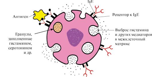 Figure: 1.33. Interaction of IgE with antigen on the mast cell surface
After the antigen attaches to the corresponding antigen-binding sites of IgE, the cells receive a signal for the secretion of biologically active substances (histamine, serotonin), which are largely responsible for the development of an inflammatory reaction and for the manifestation of allergic reactions such as asthma, urticaria, and hay fever.
Immunoglobulins D
Immunoglobulins D are found in serum in very small amounts; they are monomers. Heavy δ-chains have one variable and three constant domains. IgD acts as a receptor for B-lymphocytes, other functions are still unknown. The interaction of specific antigens with receptors on the surface of B-lymphocytes (IgD) leads to the transmission of these signals into the cell and the activation of mechanisms that ensure the multiplication of this clone of lymphocytes.
TOPIC 1.7. PHYSICO-CHEMICAL PROPERTIES OF PROTEINS AND METHODS OF THEIR SEPARATION
1. Individual proteins differ in physical and chemical properties:
The shape of the molecules;
Molecular weight;
The total charge, the value of which depends on the ratio of the anionic and cationic groups of amino acids;
The ratio of polar and non-polar amino acid radicals on the surface of molecules;
Degrees of resistance to various denaturing agents.
2. The solubility of proteins dependson the properties of the proteins listed above, as well as on the composition of the medium in which the protein is dissolved (pH, salt composition, temperature, the presence of other organic substances that can interact with the protein). The magnitude of the charge of protein molecules is one of the factors affecting their solubility. When the charge is lost at the isoelectric point, proteins aggregate more easily and precipitate. This is especially true for denatured proteins, which have hydrophobic amino acid radicals on the surface.
On the surface of a protein molecule, there are both positively and negatively charged amino acid radicals. The number of these groups, and hence the total charge of proteins, depend on the pH of the medium, i.e. the ratio of the concentration of H + - and OH - -groups. In an acidic environmentan increase in the concentration of H + leads to the suppression of the dissociation of carboxyl groups -COO - + H +\u003e - COOH and a decrease in the negative charge of proteins. In an alkaline medium, the binding of excess OH - by protons formed during the dissociation of amino groups -NH 3 + + OH - - NH 2 + H 2 O with the formation of water leads to a decrease in the positive charge of proteins. The pH value at which a protein has a total zero charge is called isoelectric point (IEP).In IEP, the number of positively and negatively charged groups is the same, i.e. the protein is in an isoelectric state.
3. Separation of individual proteins.Features of the structure and functioning of the body depend on the set of proteins synthesized in it. The study of the structure and properties of proteins is impossible without their isolation from the cell and purification from other proteins and organic molecules. Stages of isolation and purification of individual proteins:
Cell destructionof the studied tissue and obtaining a homogenate.
Separation of the homogenate into fractionscentrifugation, obtaining a nuclear, mitochondrial, cytosolic or other fraction containing the desired protein.
Selective heat denaturation- short-term heating of the protein solution, in which it is possible to remove some of the denatured protein impurities (in the event that the protein is relatively thermostable).
Salting out.Different proteins precipitate at different salt concentrations in solution. By gradually increasing the salt concentration, it is possible to obtain a number of individual fractions with a predominant content of the secreted protein in one of them. Ammonium sulfate is most commonly used for protein fractionation. Proteins with the lowest solubility precipitate at low salt concentrations.
Gel filtration- a method of sifting molecules through swollen Sephadex granules (three-dimensional polysaccharide dextran chains with pores). The rate of passage of proteins through a column filled with Sephadex will depend on their molecular weight: the smaller the mass of protein molecules, the easier they penetrate into the granules and stay there longer, the larger the mass, the faster they elute from the column.
Ultracentrifugation- a method in which proteins in a centrifuge tube are placed in an ultracentrifuge rotor. When the rotor rotates, the sedimentation rate of proteins is proportional to their molecular weight: fractions of heavier proteins are located closer to the bottom of the test tube, lighter ones are closer to the surface.
Electrophoresis- a method based on differences in the speed of movement of proteins in an electric field. This value is proportional to the charge of proteins. Protein electrophoresis is carried out on paper (in this case, the speed of movement of proteins is proportional only to their charge) or in polyacrylamide gel with a certain pore size (the speed of movement of proteins is proportional to their charge and molecular weight).
Ion exchange chromatography- a fractionation method based on the binding of ionized groups of proteins with oppositely charged groups of ion exchange resins (insoluble polymeric materials). The strength of the binding of the protein to the resin is proportional to the charge of the protein. Proteins adsorbed on an ion-exchange polymer can be washed off with NaCl solutions with increasing concentrations; the lower the charge of the protein, the lower the concentration of NaCl will be required to wash off the protein bound to the ionic groups of the resin.
Affinity chromatography- the most specific method for isolating individual proteins. A ligand of a protein is covalently attached to an inert polymer. When a solution of proteins is passed through a column with a polymer due to the complementary binding of the protein to the ligand, only the protein specific for this ligand is adsorbed on the column.
Dialysis- a method used to remove low molecular weight compounds from a solution of the isolated protein. The method is based on the inability of proteins to pass through a semipermeable membrane, in contrast to low molecular weight substances. It is used to purify proteins from low molecular weight impurities, for example, from salts after salting out.
OBJECTIVES FOR OUT-OF-AUDIT WORK
1. Fill in the table. 1.4.
Table 1.4. Comparative analysis of the structure and functions of related proteins - myoglobin and hemoglobin
a) remember the structure of the active center MB and Hb. What is the role of hydrophobic amino acid radicals in the formation of active centers of these proteins? Describe the structure of the MB and Hb active center and the mechanisms of ligand attachment to it. What role do the His F 8 and His E 7 residues play in the functioning of the MB and Hv active center?
b) what new properties in comparison with monomeric myoglobin does a closely related oligomeric protein - hemoglobin - have? Explain the role of cooperative changes in the conformation of protomers in the hemoglobin molecule, the effect of CO2 and proton concentrations on the oxygen affinity of hemoglobin, as well as the role of 2,3-BPH in the allosteric regulation of Hb function.
2. Characterize molecular chaperones, paying attention to the relationship between their structure and function.
3. What proteins are grouped into families? Using the immunoglobulin family as an example, determine the similar structural features and related functions of the proteins of this family.
4. Often purified individual proteins are required for biochemical and medical purposes. Explain on what physicochemical properties of proteins are used methods of their separation and purification.
TASKS FOR SELF-CONTROL
1. Choose the correct answers.
Hemoglobin functions:
A. Transport of O 2 from lungs to tissues B. Transport of H + from tissues to lungs
B. Maintaining a constant blood pH D. Transport of CO 2 from the lungs to tissues
E. Transport of CO 2 from tissues to lungs
2. Choose the correct answers. Ligandα -protomer HB is:A. Heme
B. Oxygen
B. CO G. 2,3-BFG
E. β-Protomer
3. Choose the correct answers.
Hemoglobin as opposed to myoglobin:
A. Has a quaternary structure
B. The secondary structure is represented only by α-helices
B. Refers to complex proteins
D. Interacts with an allosteric ligand D. Covalently linked to heme
4. Choose the correct answers.
The affinity of Нв for О 2 decreases:
A. With the addition of one O 2 molecule B. With the elimination of one O 2 molecule
B. When interacting with 2,3-BFG
D. When attached to protomers H + D. With a decrease in the concentration of 2,3-BFG
5. Establish correspondence.
Typical for HB types:
A. In deoxy form forms fibrillar aggregates B. Contains two α- and two δ-chains
B. The predominant form of HB in the erythrocytes of an adult D. In the active center contains heme with Fe + 3
E. Contains two α- and two γ-chains 1. НвА 2.
6. Establish correspondence.
Ligands HB:
A. Binds to HB in the allosteric center
B. Has a very high affinity for the active center HB
B. Joining, increases the affinity of Hb to O 2 G. Oxidizes Fe + 2 to Fe + 3
E. Forms a covalent bond with hisF8
7. Choose the correct answers.
Chaperones:
A. Proteins present in all parts of the cell
B. Synthesis is enhanced by stress
B. Participate in the hydrolysis of denatured proteins
D. Participate in maintaining the native conformation of proteins
E. Create organelles in which protein conformation is formed
8. Set the match. Immunoglobulins:
A. The secretory form is pentameric
B. Class Ig, penetrating the placental barrier
B. Ig - mast cell receptor
D. The main class of Ig present in the secretions of epithelial cells. E. B-lymphocyte receptor, the activation of which ensures cell proliferation
9. Choose the correct answers.
Immunoglobulins E:
A. Produced by macrophages B. They have heavy ε-chains.
B. Are incorporated into the T-lymphocyte membrane
D. Play the role of membrane receptors for antigens on mast cells and basophils
E. Responsible for the manifestation of allergic reactions
10. Choose the correct answers.
The method for separating proteins is based on differences in their molecular weight:
A. Gel filtration
B. Ultracentrifugation
B. Polyacrylamide gel electrophoresis D. Ion exchange chromatography
D. Affinity chromatography
11. Choose the correct answer.
The method for separating proteins is based on differences in their solubility in water:
A. Gel filtration B. Salting out
B. Ion exchange chromatography D. Affinity chromatography
D. Polyacrylamide gel electrophoresis
STANDARDS OF ANSWERS TO "TASKS FOR SELF-CONTROL"
1.A, B, C, D
2.A, B, C, D
5.1-B, 2-A, 3-D
6.1-B, 2-B, 3-A
7.A, B, D, D
8.1-D; 2-B, 3-C
BASIC TERMS AND CONCEPTS
1. Oligomeric proteins, protomere, quaternary structure of proteins
2. Cooperative changes in the conformation of protomers
3. Bohr effect
4. Allosteric regulation of protein functions, allosteric center and allosteric effector
5. Molecular chaperones, heat shock proteins
6. Protein families (serine proteases, immunoglobulins)
7. IgM-, G-, E-, A-connection of structure with function
8. The total charge of proteins, isoelectric point of proteins
9. Electrophoresis
10. Salting out
11. Gel filtration
12. Ion exchange chromatography
13. Ultracentrifugation
14. Affinity chromatography
15. Electrophoresis of blood plasma proteins
TASKS FOR AUDITING
1. Compare the dependences of the degrees of saturation of hemoglobin (Hb) and myoglobin (Mb) with oxygen on its partial pressure in the tissues
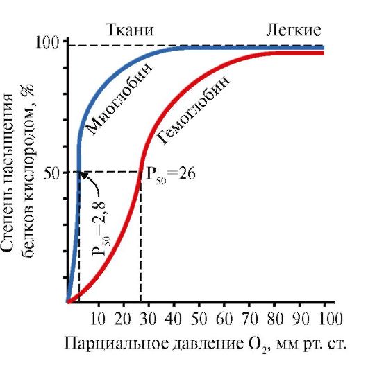 Figure: 1.34. Saturation dependence of MV andHboxygen from its partial pressure
Note that the shape of the curves of protein oxygen saturation is different: for myoglobin - hyperbole, for hemoglobin - sigmoid shape.
1.Compare the values \u200b\u200bof the partial pressure of oxygen, at which MB and HB are saturated with O 2 by 50%. Which of these proteins has a higher affinity for O 2?
2. What features of the structure of MB determine its high affinity for О 2?
3. What features of the structure of HB allow it to release O 2 in the capillaries of resting tissues (at a relatively high partial pressure of O 2) and sharply increase this return in working muscles? What property of oligomeric proteins provides this effect?
4.Calculate what amount of O 2 (in%) gives oxygenated hemoglobin to a resting and working muscle?
5. draw conclusions about the relationship between the structure of the protein and its function.
2. The amount of oxygen released by hemoglobin in capillaries depends on the intensity of catabolic processes in tissues (Bohr effect). How do changes in tissue metabolism regulate the affinity of Hb for O 2? Effect of CO 2 and H + on the affinity of Hb for O 2
 1. describe the Bohr effect.
2. in which direction is the process shown in the diagram:
a) in the capillaries of the lungs;
b) in tissue capillaries?
3. What is the physiological significance of the Bohr effect?
4. Why does the interaction of Hb with H + at sites remote from the heme change the affinity of the protein for O 2?
3. The affinity of Hb for O 2 depends on the concentration of its ligand, 2,3-bisphosphoglycerate, which is an allosteric regulator of the affinity of Hb for O 2. Why does the interaction of a ligand at a site distant from the active site affect protein function? How does 2,3-BFG regulate the affinity of Hb for O 2? To solve the problem, answer the following questions:
1.Where and from what is 2.3-bisphosphoglycerate (2,3-BFG) synthesized? Write down its formula, indicate the charge of a given molecule.
2. What form of hemoglobin (oxy or deoxy) does BFG interact with and why? In what part of the HB molecule does the interaction take place?
3. in which direction is the process shown in the diagram
a) in tissue capillaries;
b) in the capillaries of the lungs?
4.where the concentration of the complex should be higher
NV-2,3-BFG:
a) in the capillaries of muscles at rest,
b) in the capillaries of working muscles (assuming the same concentration of BFG in erythrocytes)?
5. How will the affinity of Hb for oxygen change when a person adapts to high altitude conditions, if the concentration of BFG in erythrocytes increases? What is the physiological significance of this phenomenon?
4. Destruction of 2,3-BFG during storage of canned blood disrupts the function of HB. How will the affinity of HB to O 2 in preserved blood change if the concentration of 2,3-BFG in erythrocytes can decrease from 8 to 0.5 mmol / l. Is it possible to transfuse such blood to critically ill patients if the concentration of 2,3-BFG is restored no earlier than three days later? Is it possible to restore the function of erythrocytes by adding 2,3-BFG to the blood?
5. Remember the structure of the simplest molecules of immunoglobulins. What is the role of immunoglobulins in the immune system? Why are Ig often called bivalents? How is the structure of Ig related to their function? (Describe an example of a class of immunoglobulins.)
Physicochemical properties of proteins and methods of their separation.
6. How does the net charge of a protein affect its solubility?
a) determine the total charge of the peptide at pH 7
Ala-Glu-Tre-Pro-Asp-Liz-Cis
b) how the charge of this peptide will change at pH\u003e 7, pH<7, рН <<7?
c) what is the isoelectric point of a protein (IEP) and in what environment does it lie
IEP of this peptide?
d) at what pH value the least solubility of this peptide will be observed.
7. Why does sour milk, in contrast to fresh milk, "curdle" when boiled (ie, casein milk protein precipitates)? In fresh milk, casein molecules have a negative charge.
8. Gel filtration is used to separate individual proteins. A mixture containing proteins A, B, C with molecular weights of 160,000, 80,000 and 60,000, respectively, was analyzed by gel filtration (Fig. 1.35). The swollen gel granules are permeable to proteins with a molecular weight of less than 70,000. What is the principle behind this separation method? Which graph correctly represents the fractionation results? Specify the order in which proteins A, B, and C come out of the column.
 Figure: 1.35. Using a gel filtration method to separate proteins
9. In fig. 1.36, A shows a diagram of electrophoresis on paper of blood serum proteins of a healthy person. The relative amounts of protein fractions obtained using this method are: albumin 54-58%, α 1 -globulins 6-7%, α 2 -globulins 8-9%, β-globulins 13%, γ-globulins 11-12% ...
 Figure: 1.36 Electrophoresis on paper of blood plasma proteins of a healthy person (A) and a patient (B)
I - γ-globulins; II - β-globulins; III -α 2 -globulin; IV -α 2 -globulin; V - albumin
Many diseases are accompanied by quantitative changes in the composition of serum proteins (dysproteinemia). The nature of these changes is taken into account when making a diagnosis and assessing the severity and stage of the disease.
Using the data shown in table. 1.5, make an assumption about the disease, which is characterized by the electrophoretic profile shown in Fig. 1.36.
Table 1.5. Changes in the concentration of serum proteins in pathology
Module structure | Topics |
Modular unit 1 | 1.1. Structural organization of proteins. Stages of the formation of native protein conformation 1.2. Fundamentals of protein functioning. Drugs as ligands that affect protein function 1.3. Denaturation of proteins and the possibility of their spontaneous renewal |
Modular unit 2 | 1.4. Features of the structure and functioning of oligomeric proteins on the example of hemoglobin 1.5. Maintaining native conformation of proteins under cell conditions 1.6. Variety of proteins. Protein families as exemplified by immunoglobulins 1.7. Physicochemical properties of proteins and methods of their separation |
Modular unit 1 STRUCTURAL ORGANIZATION OF MONOMERIC PROTEINS AND THE BASIS OF THEIR FUNCTIONING
Learning Objectives To be able to:
1. Use knowledge about the features of the structure of proteins and the dependence of the functions of proteins on their structure to understand the mechanisms of development of hereditary and acquired proteinopathies.
2. Explain the mechanisms of the therapeutic action of some drugs as ligands that interact with proteins and change their activity.
3. Use knowledge of the structure and conformational lability of proteins to understand their structural and functional instability and tendency to denaturation under changing conditions.
4. Explain the use of denaturing agents as means for sterilizing medical material and instruments, as well as antiseptics.
Know:
1. Levels of the structural organization of proteins.
2. The value of the primary structure of proteins, which determines their structural and functional diversity.
3. The mechanism of formation of an active center in proteins and its specific interaction with the ligand, which underlies the functioning of proteins.
4. Examples of the influence of exogenous ligands (drugs, toxins, poisons) on the conformation and functional activity of proteins.
5. Causes and consequences of protein denaturation, factors causing denaturation.
6. Examples of the use of denaturing factors in medicine as antiseptics and means for sterilizing medical instruments.
TOPIC 1.1. STRUCTURAL ORGANIZATION OF PROTEINS. STAGES OF FORMATION OF NATIVE
PROTEIN CONFORMATIONS
Proteins are polymer molecules with a total of 20 α-amino acids as monomers. The set and order in which amino acids are combined in a protein is determined by the structure of genes in the DNA of individuals. Each protein, in accordance with its specific structure, performs its own function. The set of proteins of a given organism determines its phenotypic characteristics, as well as the presence of hereditary diseases or a predisposition to their development.
1. Amino acids that make up proteins. Peptide bond.Proteins are polymers built from monomers - 20 α-amino acids, the general formula of which is
Amino acids differ in structure, size, physicochemical properties of the radicals attached to the α-carbon atom. The functional groups of amino acids determine the characteristics of the properties of different α-amino acids. The radicals found in α-amino acids can be divided into several groups:
Proline,unlike other 19 protein monomers, it is not an amino acid, but an imino acid, the radical in proline is linked to both the α-carbon atom and the imino group
Amino acids differ in their solubility in water.This is due to the ability of radicals to interact with water (hydrate).
TO hydrophilicincludes radicals containing anionic, cationic and polar uncharged functional groups.
TO hydrophobicincludes radicals containing methyl groups, aliphatic chains or cycles.
2. Peptide bonds link amino acids into peptides.In the synthesis of a peptide, the α-carboxyl group of one amino acid interacts with the α-amino group of another amino acid to form peptide bond:
Proteins are polypeptides, i.e. linear polymers of α-amino acids linked by a peptide bond (Fig.1.1.)
Figure: 1.1. Terms used to describe the structure of peptides
Monomers of amino acids that make up polypeptides are called amino acid residues.A chain of repeating groups - NH-CH-CO- forms peptide backbone.An amino acid residue that has a free α-amino group is called N-terminal, and one that has a free α-carboxyl group is called C-terminal. The peptides are written and read from the N-terminus to the C-terminus.
The peptide bond formed by the imino group of proline differs from other peptide bonds: the nitrogen atom of the peptide group lacks hydrogen,
instead, there is a bond with the radical, as a result, one side of the cycle is included in the peptide backbone:
Peptides differ in amino acid composition, the number of amino acids and the order in which the amino acids are combined, for example, Ser-Ala-Glu-Gis and Gis-Glu-Ala-Ser are two different peptides.
Peptide bonds are very strong, and strict conditions are required for their chemical non-enzymatic hydrolysis: the analyzed protein is hydrolyzed in concentrated hydrochloric acid at a temperature of about 110 ° for 24 hours. In a living cell, peptide bonds can be broken using proteolytic enzymes,called proteasesor peptide hydrolases.
3. Primary structure of proteins.Amino acid residues in peptide chains of different proteins do not alternate randomly, but are arranged in a certain order. The linear sequence or the order of alternation of amino acid residues in the polypeptide chain is called the primary structure of the protein.
The primary structure of each individual protein is encoded in a DNA molecule (in a region called a genome) and is realized during transcription (rewriting information on mRNA) and translation (synthesis of the primary structure of a protein). Consequently, the primary structure of proteins of an individual person is information hereditarily transmitted from parents to children, which determines the structural features of the proteins of a given organism, on which the function of existing proteins depends (Fig. 1.2.).
Figure: 1.2. The relationship between the genotype and the conformation of proteins synthesized in the body of an individual
Each of the approximately 100,000 individual proteins in the human body has uniqueprimary structure. The molecules of one type of protein (for example, albumin) have the same alternation of amino acid residues, which distinguishes albumin from any other individual protein.
The sequence of amino acid residues in the peptide chain can be considered as a form of recording information. This information determines the spatial folding of the linear peptide chain into a more compact three-dimensional structure called conformationsquirrel. The process of forming a functionally active protein conformation is called folding.
4. Conformation of proteins.Free rotation in the peptide backbone is possible between the nitrogen atom of the peptide group and the adjacent α-carbon atom, as well as between the α-carbon atom and the carbonyl carbon. Due to the interaction of functional groups of amino acid residues, the primary structure of proteins can acquire more complex spatial structures. In globular proteins, there are two main levels of folding of the conformation of peptide chains: secondaryand tertiary structure.
Secondary structure of proteinsis a spatial structure formed as a result of the formation of hydrogen bonds between the functional groups -C \u003d O and - NH- of the peptide backbone. In this case, the peptide chain can acquire regular structures of two types: α-helixand β-structures.
IN α-helixhydrogen bonds are formed between the oxygen atom of the carbonyl group and the hydrogen of the amide nitrogen of the 4th amino acid from it; side chains of amino acid residues
are located along the periphery of the spiral, without participating in the formation of the secondary structure (Fig. 1.3.).
Bulky radicals or radicals carrying the same charges prevent the formation of the α-helix. The proline residue, which has a ring structure, interrupts the α-helix, since a hydrogen bond cannot be formed due to the absence of hydrogen at the nitrogen atom in the peptide chain. The bond between nitrogen and the α-carbon atom is part of the proline cycle; therefore, the peptide backbone at this point acquires a bend.
β-structureforms between the linear regions of the peptide backbone of one polypeptide chain, thus forming folded structures. Polypeptide chains or parts thereof can form parallelor antiparallel β-structures.In the first case, the N- and C-ends of the interacting peptide chains coincide, and in the second, they have the opposite direction (Fig. 1.4).
Figure: 1.3. Secondary structure of protein - α-helix
Figure: 1.4. Parallel and antiparallel β-fold structures
β-structures are indicated by wide arrows: A - Antiparallel β-structure. B - Parallel β-folded structures
In some proteins, β-structures can be formed due to the formation of hydrogen bonds between the atoms of the peptide backbone of different polypeptide chains.
Also found in proteins areas with irregular secondarystructure, which include bends, loops, turns of the polypeptide backbone. They are often located in places where the direction of the peptide chain changes, for example, when a parallel β-sheet structure is formed.
According to the presence of α-helices and β-structures, globular proteins can be divided into four categories.
Figure: 1.5. Secondary structure of myoglobin (A) and β-chains of hemoglobin (B), containing eight α-helices
Figure: 1.6. Secondary structure of triose phosphate isomerase and pyruvate kinase domain
Figure: 1.7. Secondary structure of the constant domain of immunoglobulin (A) and the enzyme superoxide dismutase (B)
IN fourth categoryincluded proteins containing a small amount of regular secondary structures. These proteins include small, cysteine-rich proteins or metalloproteins.
Protein tertiary structure- the type of conformation formed due to interactions between amino acid radicals, which can be located at a considerable distance from each other in the peptide chain. In this case, most proteins form a spatial structure resembling a globule (globular proteins).
Since hydrophobic amino acid radicals tend to combine with the so-called hydrophobic interactionsand intermolecular van der Waals forces, a dense hydrophobic core is formed inside the protein globule. Hydrophilic ionized and non-ionized radicals are mainly located on the surface of the protein and determine its solubility in water.
Figure: 1.8. Types of bonds that arise between amino acid radicals during the formation of the tertiary structure of a protein
1 - ionic bond- occurs between positively and negatively charged functional groups;
2 - hydrogen bond- occurs between an uncharged hydrophilic group and any other hydrophilic group;
3 - hydrophobic interactions- arise between hydrophobic radicals;
4 - disulfide bond- formed due to the oxidation of SH-groups of cysteine \u200b\u200bresidues and their interaction with each other
Hydrophilic amino acid residues trapped inside the hydrophobic core can interact with each other using ionicand hydrogen bonds(fig. 1.8).
Ionic and hydrogen bonds, as well as hydrophobic interactions, are among the weak ones: their energy is not much higher than the energy of the thermal motion of molecules at room temperature. Protein conformation is maintained through the emergence of many of these weak bonds. Since the atoms that make up the protein are in constant motion, some weak bonds can be broken and others formed, which leads to small displacements of individual sections of the polypeptide chain. This property of proteins to change conformation as a result of breaking some and the formation of other weak bonds is called conformational lability.
The human body has systems that support homeostasis- the constancy of the internal environment within certain limits permissible for a healthy organism. Under homeostasis conditions, small changes in conformation do not disrupt the overall structure and function of proteins. The functionally active conformation of a protein is called native conformation.A change in the internal environment (for example, the concentration of glucose, Ca ions, protons, etc.) leads to a change in conformation and dysfunction of proteins.
The tertiary structure of some proteins is stabilized disulfide bonds,formed due to the interaction of -SH groups of two residues
Figure: 1.9. Formation of a disulfide bond in a protein molecule
cysteine \u200b\u200b(Fig. 1.9). Most intracellular proteins do not have covalent disulfide bonds in the tertiary structure. Their presence is characteristic of proteins secreted by the cell, which ensures their greater stability in extracellular conditions. So, disulfide bonds are found in insulin and immunoglobulin molecules.
Insulin- a protein hormone synthesized in the β-cells of the pancreas and secreted into the blood in response to an increase in the concentration of glucose in the blood. In the structure of insulin, there are two disulfide bonds connecting the polypeptide A- and B-chains, and one disulfide bond within the A-chain (Fig. 1.10).
Figure: 1.10. Disulfide bonds in the structure of insulin
5. Supersecondary structure of proteins.In proteins that differ in their primary structure and functions, sometimes similar combinations and arrangement of secondary structures,which are called supersecondary structure. It occupies an intermediate position between the secondary and tertiary structures, since this is a specific combination of elements of the secondary structure in the formation of the tertiary structure of the protein. Supersecondary structures have specific names such as “α-helix-turn-a-helix”, “leucine zipper”, “zinc fingers”, etc. Such supersecondary structures are characteristic of DNA-binding proteins.
Leucine zipper.This kind of supersecondary structure is used to connect two proteins. On the surface of interacting proteins, there are α-helical regions containing at least four leucine residues. Leucine residues in the α-helix are located six amino acids apart from each other. Since each turn of the α-helix contains 3.6 amino acid residues, leucine radicals are located on the surface of every second turn. Leucine residues of the α-helix of one protein can interact with leucine residues of another protein (hydrophobic interactions), linking them together (Fig. 1.11.). Many DNA-binding proteins function as part of oligomeric complexes, where individual subunits bind to each other with "leucine fasteners".
Figure: 1.11. "Leucine zipper" between the α-helical regions of two proteins
An example of such proteins is histones. Histones- nuclear proteins, which include a large number of positively charged amino acids - arginine and lysine (up to 80%). Histone molecules are combined into oligomeric complexes containing eight monomers with the help of "leucine fasteners", despite the significant charge of the same name of these molecules.
"Zinc finger"- a variant of the supersecondary structure, characteristic of DNA-binding proteins, has the form of an elongated fragment on the protein surface and contains about 20 amino acid residues (Fig. 1.12). The shape of the "elongated finger" is supported by a zinc atom linked to the radicals of four amino acids - two cysteine \u200b\u200bresidues and two histidine residues. In some cases, instead of histidine residues, there are cysteine \u200b\u200bresidues. Two closely spaced cysteine \u200b\u200bresidues are separated from the other two Gisili residues by a sequence of about 12 amino acid residues. This region of the protein forms an α-helix, the radicals of which can specifically bind to the regulatory regions of the major groove of DNA. The binding specificity of an individual
Figure: 1.12. The primary structure of a region of DNA-binding proteins that form the structure of the "zinc finger" (letters indicate the amino acids that make up this structure)
regulatory DNA-binding protein depends on the sequence of amino acid residues located in the "zinc finger". Such structures contain, in particular, receptors for steroid hormones involved in the regulation of transcription (reading information from DNA to RNA).
TOPIC 1.2. BASES OF FUNCTIONING OF PROTEINS. MEDICINES AS LIGANDS AFFECTING THE FUNCTION OF PROTEINS
1. The active center of the protein and its interaction with the ligand.During the formation of a tertiary structure on the surface of a functionally active protein, usually in a depression, a region is formed that is formed by amino acid radicals that are far from each other in the primary structure. This site, which has a unique structure for a given protein and is able to specifically interact with a certain molecule or a group of similar molecules, is called the protein-ligand binding site or active site. Ligands are molecules that interact with proteins.
High specificitythe interaction of the protein with the ligand is provided by the complementarity of the structure of the active center to the structure of the ligand.
Complementarityis the spatial and chemical correspondence of interacting surfaces. The active center should not only spatially correspond to the ligand included in it, but also bonds (ionic, hydrogen, and also hydrophobic interactions) should form between the functional groups of the radicals included in the active center and the ligand, which hold the ligand in the active center (Fig.1.13 ).
Figure: 1.13. Complementary interaction of a protein with a ligand
Some ligands, by attaching to the active center of the protein, play an auxiliary role in the functioning of proteins. Such ligands are called cofactors, and proteins containing a non-protein part are called complex proteins(as opposed to simple proteins, consisting only of the protein part). The non-protein part, firmly attached to the protein, is called prosthetic group.For example, the composition of myoglobin, hemoglobin and cytochromes contains a prosthetic group firmly attached to the active center - heme, which contains an iron ion. The complex proteins that contain heme are called hemoproteins.
When specific ligands are attached to proteins, the function of these proteins is manifested. So, albumin - the most important protein in blood plasma - manifests its transport function by attaching hydrophobic ligands such as fatty acids, bilirubin, some drugs, etc. to the active center (Fig.1.14)
Ligands interacting with the three-dimensional structure of the peptide chain can be not only low molecular weight organic and inorganic molecules, but also macromolecules:
DNA (examples of DNA-binding proteins discussed above);
Polysaccharides;
Figure: 1.14. Relationship between genotype and phenotype
The unique primary structure of human proteins, encoded in the DNA molecule, is realized in cells in the form of a unique conformation, structure of the active center and functions of proteins
In these cases, the protein recognizes a certain ligand site commensurate with and complementary to the binding site. So on the surface of hepatocytes there are receptor proteins for the hormone insulin, which also has a protein structure. The interaction of insulin with the receptor causes a change in its conformation and activation of signaling systems, leading to the accumulation of nutrients in hepatocytes after eating.
Thus, the functioning of proteins is based on the specific interaction of the active center of the protein with the ligand.
2. Domain structure and its role in the functioning of proteins.Long polypeptide chains of globular proteins often fold into several compact, relatively independent regions. They have an independent tertiary structure, similar to that of globular proteins, and are called domains.Due to the domain structure of proteins, their tertiary structure is more easily formed.
In domain proteins, ligand binding sites are often located between domains. Thus, trypsin is a proteolytic enzyme that is produced by the exocrine part of the pancreas and is necessary for the digestion of food proteins. It has a two-domain structure, and the binding site of trypsin with its ligand, a food protein, is located in the groove between the two domains. In the active center, conditions are created that are necessary for the effective binding of a specific site of the food protein and the hydrolysis of its peptide bonds.
When the active center interacts with the ligand, different domains in a protein can move relative to each other (Fig. 1.15).
Hexokinase- an enzyme that catalyzes the phosphorylation of glucose by means of ATP. The active site of the enzyme is located in the cleft between the two domains. When hexokinase binds to glucose, the surrounding domains close and the substrate is trapped, where phosphorylation occurs (see Fig. 1.15).
Figure: 1.15. Binding of Hexokinase Domains to Glucose
In some proteins, domains perform independent functions by binding to various ligands. These proteins are called multifunctional.
3. Drugs - ligands that affect the function of proteins.The interaction of proteins with ligands is specific. However, due to the conformational lability of the protein and its active center, it is possible to select another substance that could also interact with the protein in the active center or other part of the molecule.
A substance similar in structure to a natural ligand is called structural analogue of the ligandor an unnatural ligand. It also interacts with a protein in the active site. The structural analogue of the ligand can both enhance the function of the protein (agonist),so reduce it (antagonist).The ligand and its structural analogs compete with each other for binding to the protein at the same center. Such substances are called competitive modulators(regulators) of protein functions. Many drugs act as protein inhibitors. Some of them are obtained by chemical modification of natural ligands. Protein inhibitors can be drugs and poisons.
Atropine is a competitive inhibitor of M-cholinergic receptors.Acetylcholine is a neurotransmitter for the transmission of nerve impulses through cholinergic synapses. To conduct excitation, acetylcholine released into the synaptic cleft must interact with a protein - the receptor of the postsynaptic membrane. Two types found cholinergic receptors:
M-receptor,in addition to acetylcholine, it selectively interacts with muscarin (fly agaric toxin). M - cholinergic receptors are present on smooth muscles and, when interacting with acetylcholine, cause their contraction;
H-receptor,specifically binding to nicotine. H-cholinergic receptors are found in the synapses of striated skeletal muscles.
Specific inhibitor M-cholinergic receptorsis atropine. It is found in belladonna and henbane plants.
Atropine has in the structure functional groups similar to acetylcholine and their spatial arrangement, therefore, it belongs to competitive inhibitors of M-cholinergic receptors. Considering that the binding of acetylcholine to M-cholinergic receptors causes contraction of smooth muscles, atropine is used as a medicine that relieves muscle spasms. (antispasmodic).So, the use of atropine is known to relax the eye muscles when looking at the fundus, as well as to relieve spasms in gastrointestinal colic. M-cholinergic receptors are also present in the central nervous system (CNS), therefore, large doses of atropine can cause an unwanted reaction from the central nervous system: motor and mental agitation, hallucinations, convulsions.
Ditilin is a competitive agonist of H-cholinergic receptors that inhibits the function of neuromuscular synapses.
Skeletal muscle neuromuscular synapses contain H-cholinergic receptors. Their interaction with acetylcholine leads to muscle contractions. Some surgical procedures, as well as endoscopic examinations, use drugs that cause relaxation of skeletal muscles (muscle relaxants).These include dityline, which is a structural analog of acetylcholine. It attaches to H-cholinergic receptors, but unlike acetylcholine, it is very slowly destroyed by the enzyme, acetylcholinesterase. As a result of prolonged opening of ion channels and persistent depolarization of the membrane, the conduction of the nerve impulse is disrupted and muscle relaxation occurs. Initially, these properties were found in curare poison, therefore such drugs are called curariform.
TOPIC 1.3. DENATURATION OF PROTEINS AND THE POSSIBILITY OF THEIR SPONTANEOUS RENATIVATION
1. Since the native conformation of proteins is maintained due to weak interactions, changes in the composition and properties of the environment surrounding the protein, exposure to chemical reagents and physical factors cause a change in their conformation (property of conformational lability). The breaking of a large number of bonds leads to the destruction of the native conformation and denaturation of proteins.
Protein denaturationis the destruction of their native conformation under the action of denaturing agents, caused by the breaking of weak bonds that stabilize the spatial structure of the protein. Denaturation is accompanied by the destruction of the unique three-dimensional structure and active center of the protein and the loss of its biological activity (Fig. 1.16).
All denatured molecules of one protein acquire a random conformation that differs from other molecules of the same protein. The amino acid radicals that form the active center are spatially distant from each other, i.e. the specific binding site of the protein with the ligand is destroyed. During denaturation, the primary structure of proteins remains unchanged.
The use of denaturing agents in biological research and medicine.In biochemical studies, before the determination of low molecular weight compounds in biological material, proteins are usually removed from the solution first. For this purpose, trichloroacetic acid (TCA) is most often used. After adding TCA to the solution, denatured proteins precipitate and are easily removed by filtration (Table 1.1.)
In medicine, denaturing agents are often used to sterilize medical instruments and materials in autoclaves (denaturing agent - high temperature) and as antiseptics (alcohol, phenol, chloramine) to treat contaminated surfaces containing pathogenic microflora.
2. Spontaneous protein renewal- proof of the determinism of the primary structure, conformation and function of proteins. Individual proteins are products of one gene that have an identical amino acid sequence and acquire the same conformation in the cell. The fundamental conclusion that the primary structure of a protein already contains information about its conformation and function was made on the basis of the ability of some proteins (in particular, ribonuclease and myoglobin) to spontaneously regenerate - to restore their native conformation after denaturation.
The formation of spatial structures of a protein is carried out by the method of self-assembly - a spontaneous process in which a polypeptide chain, which has a unique primary structure, tends to accept a conformation with the lowest free energy in solution. The ability to renew proteins that retain their primary structure after denaturation was described in an experiment with the enzyme ribonuclease.
Ribonuclease is an enzyme that breaks down the bonds between individual nucleotides in an RNA molecule. This globular protein has a single polypeptide chain, the tertiary structure of which is stabilized by many weak and four disulfide bonds.
Treatment of ribonuclease with urea, which destroys hydrogen bonds in the molecule, and a reducing agent that breaks disulfide bonds, leads to denaturation of the enzyme and the loss of its activity.
Removal of denaturing agents by dialysis results in restoration of protein conformation and function, i.e. to the renewal. (fig. 1.17).
Figure: 1.17. Denaturation and renaissance of ribonuclease
A - native conformation of ribonuclease, in the tertiary structure of which there are four disulfide bonds; B - denatured ribonuclease molecule;
B - revived ribonuclease molecule with reduced structure and function
1. Complete table 1.2.
Table 1.2. Classification of amino acids by radical polarity
2. Write the formula for tetrapeptide:
Asp - Pro - Fen - Liz
a) isolate in the peptide the repeating groups that form the peptide backbone and variable groups represented by amino acid radicals;
b) designate the N- and C-ends;
c) underline peptide bonds;
d) write another peptide consisting of the same amino acids;
e) count the number of possible variants of a tetrapeptide with a similar amino acid composition.
3. Explain the role of the primary structure of proteins by the example of a comparative analysis of two structurally similar and evolutionarily close peptide hormones of the mammalian neurohypophysis - oxytocin and vasopressin (Table 1.3).
Table 1.3. Structure and function of oxytocin and vasopressin
For this:
a) compare the composition and amino acid sequence of the two peptides;
b) find the similarity of the primary structure of two peptides and the similarity of their biological action;
c) find the differences in the structure of the two peptides and the difference in their functions;
d) draw a conclusion about the influence of the primary structure of peptides on their functions.
4. Describe the main stages in the formation of the conformation of globular proteins (secondary, tertiary structures, the concept of a supersecondary structure). Indicate the types of bonds involved in the formation of protein structures. Which amino acid radicals can participate in the formation of hydrophobic interactions, ionic, hydrogen bonds.
Give examples.
5. Give a definition of the concept of "conformational lability of proteins", indicate the reasons for its existence and significance.
6. Expand the meaning of the following phrase: "The functioning of proteins is based on their specific interaction with the ligand", using the terms and explaining their meaning: protein conformation, active center, ligand, complementarity, protein function.
7. Use one example to explain what domains are and what their role in proteins is.
TASKS FOR SELF-CONTROL
1. Establish correspondence.
Functional group in the amino acid radical:
A. Carboxyl group B. Hydroxyl group C Guanidine group D. Thiol group D. Amino group
2. Choose the correct answers.
Amino acids with polar uncharged radicals are:
A. Cis B. Asn
B. Glu G. Three
3. Choose the correct answers.
Amino acid radicals:
A. Provide specificity of the primary structure B. Participate in the formation of the tertiary structure
B. Located on the surface of the protein, affect its solubility D. Form an active center
E. Participate in the formation of peptide bonds
4. Choose the correct answers.
Hydrophobic interactions can form between amino acid radicals:
A. Tre Lei B. Pro Three
B. Met Ile G. Tir Ala D. Val Fen
5. Choose the correct answers.
Ionic bonds can form between amino acid radicals:
A. Gln Asp B. Apr Liz
B. Liz Glu G. Gies Asp D. Asn Apr
6. Choose the correct answers.
Hydrogen bonds can form between amino acid radicals:
A. Ser Gln B. Cis Tre
B. Asp Liz G. Glu Asp D. Asn Tre
7. Establish correspondence.
The type of bond involved in the formation of the protein structure:
A. Primary structure B. Secondary structure
B. Tertiary structure
D. Supersecondary structure E. Conformation.
1. Hydrogen bonds between the atoms of the peptide backbone
2. Weak bonds between functional groups of amino acid radicals
3. Relationships between α-amino and α-carboxyl groups of amino acids
8. Choose the correct answers. Trypsin:
A. Proteolytic enzyme B. Contains two domains
B. Hydrolyzes starch
D. The active center is located between the domains. D. Consists of two polypeptide chains.
9. Choose the correct answers. Atropine:
A. Neurotransmitter
B. Structural analogue of acetylcholine
B. Interacts with H-cholinergic receptors
D. Strengthens the conduction of nerve impulses through cholinergic synapses
D. Competitive inhibitor of M-cholinergic receptors
10. Choose the correct statements. In proteins:
A. Primary structure contains information about the structure of its active center
B. The active center is formed at the level of the primary structure
B. The conformation is rigidly fixed by covalent bonds
D. The active site can interact with a group of similar ligands
due to the conformational lability of proteins D. Changes in the environment can affect the affinity of the active
center to ligand
1.1-C, 2-D, 3-B.
3.A, B, C, D.
7.1-B, 2-D, 3-A.
8.A, B, C, D.
BASIC TERMS AND CONCEPTS
1. Protein, polypeptide, amino acids
2. Primary, secondary, tertiary protein structure
3. Conformation, native protein conformation
4. Covalent and weak bonds in a protein
5. Conformational lability
6. Active center of protein
7. Ligands
8. Protein folding
9. Structural analogs of ligands
10. Domain proteins
11. Simple and complex proteins
12. Protein denaturation, denaturing agents
13. Renovation of proteins
Solve tasks
"Structural organization of proteins and the basis of their functioning"
1. The main function of the protein, hemoglobin A (HbA), is to transport oxygen to tissues. In the human population, multiple forms of this protein with altered properties and functions are known - the so-called abnormal hemoglobins. For example, it was found that hemoglobin S, found in the erythrocytes of sickle cell anemia (HbS) patients, has low solubility under conditions of low oxygen partial pressure (as is the case in venous blood). This leads to the formation of aggregates of this protein. The protein loses its function, precipitates, and red blood cells acquire an irregular shape (some of them form a sickle) and are destroyed faster than usual in the spleen. As a result, sickle cell anemia develops.
The only difference in the primary structure of HbA was found in the N-terminal region of the β-chain of hemoglobin. Compare the N-terminus of the β-chain and show how changes in the primary structure of a protein affect its properties and functions.
For this:
a) write the amino acid formulas by which HBA differ and compare the properties of these amino acids (polarity, charge).
b) draw a conclusion about the reason for the decrease in solubility and impaired oxygen transport in the tissue.
2. The figure shows a schematic diagram of the structure of a protein that has a ligand-binding site (active site). Explain why a protein is selective in its choice of ligand. For this:
a) remember what the active center of a protein is, and consider the structure of the active center of the protein shown in the figure;
b) write the formulas of amino acid radicals that make up the active center;
c) draw a ligand that could specifically interact with the active center of the protein. Indicate on it the functional groups capable of forming bonds with the amino acid radicals that make up the active center;
d) indicate the types of bonds that arise between the ligand and the amino acid radicals of the active center;
e) explain what the specificity of the interaction of the protein with the ligand is based on.
3.
The figure shows the active site of the protein and several ligands.
Determine which ligand is most likely to interact with the active site of the protein and why.
What types of bonds arise during the formation of a protein-ligand complex?
4. Structural analogs of natural protein ligands can be used as drugs to alter the activity of proteins.
Acetylcholine is a transmitter of excitation in neuromuscular synapses. When acetylcholine interacts with proteins - receptors of the postsynaptic membrane of skeletal muscles, ion channels open and muscle contraction. Ditilin is a medicine used in some operations to relax muscles, since it disrupts the transmission of nerve impulses through the neuromuscular synapses. Explain the mechanism of action of ditilin as a muscle relaxant drug. For this:
a) write the formulas of acetylcholine and ditilin and compare their structures;
b) describe the mechanism of the relaxing action of ditilin.
5. In some diseases, the patient's body temperature rises, which is considered as a protective reaction of the body. However, high temperatures are detrimental to body proteins. Explain why at temperatures above 40 ° C, the function of proteins is disrupted and a threat to human life arises. To do this, remember:
1) The structure of proteins and bonds that hold its structure in the native conformation;
2) How does the structure and function of proteins change with increasing temperature?;
3) What is homeostasis and why is it important for maintaining human health.
Modular unit 2 OLIGOMERIC PROTEINS AS TARGETS OF REGULATORY IMPACT. STRUCTURAL AND FUNCTIONAL PROTEIN VARIETY. PROTEIN SEPARATION AND PURIFICATION METHODS
Learning Objectives To be able to:
1. Use knowledge about the features of the structure and functions of oligomeric proteins to understand the adaptive mechanisms of regulation of their functions.
2. Explain the role of chaperones in the synthesis and maintenance of protein conformation under cell conditions.
3. Explain the variety of manifestations of life by the variety of structures and functions of proteins synthesized in the body.
4. To analyze the relationship between the structure of proteins and their function by examples of comparing related hemoproteins - myoglobin and hemoglobin, as well as representatives of five classes of proteins of the immunoglobulin family.
5. Apply knowledge about the peculiarities of the physicochemical properties of proteins to select methods for their purification from other proteins and impurities.
6. Interpret the results of the quantitative and qualitative composition of blood plasma proteins to confirm or clarify the clinical diagnosis.
Know:
1. Features of the structure of oligomeric proteins and adaptive mechanisms of regulation of their functions on the example of hemoglobin.
2. The structure and function of chaperones and their importance for maintaining the native conformation of proteins under cell conditions.
3. Principles of combining proteins into families according to the similarity of their conformation and functions on the example of immunoglobulins.
4. Methods for the separation of proteins based on the peculiarities of their physicochemical properties.
5. Electrophoresis of blood plasma as a method for assessing the qualitative and quantitative composition of proteins.
TOPIC 1.4. FEATURES OF THE STRUCTURE AND FUNCTIONING OF OLIGOMERIC PROTEINS ON THE EXAMPLE OF HEMOGLOBIN
1. Many proteins contain several polypeptide chains. Such proteins are called oligomeric,and individual chains - protomers.Protomers in oligomeric proteins are linked by many weak non-covalent bonds (hydrophobic, ionic, hydrogen). Interaction
protomers is carried out thanks complementaritytheir contact surfaces.
The number of protomers in oligomeric proteins can vary greatly: hemoglobin contains 4 protomers, the aspartate aminotransferase enzyme contains 12 protomers, and the tobacco mosaic virus protein contains 2,120 protomers linked by non-covalent bonds. Consequently, oligomeric proteins can have very high molecular weights.
The interaction of one protomer with others can be considered as a special case of the interaction of a protein with a ligand, since each protomer serves as a ligand for other protomers. The number and method of combining protomers in a protein is called quaternary protein structure.
Proteins can include protomers of the same or different structure, for example, homodimers are proteins containing two identical protomers, and heterodimers are proteins containing two different protomers.
If proteins contain different protomers, then different structures of binding sites with different ligands can be formed on them. When the ligand binds to the active site, the function of this protein is manifested. The center located on another protomer is called allosteric (other than the active one). By contacting an allosteric ligand or effector,it performs a regulatory function (Fig. 1.18). The interaction of the allosteric center with the effector causes conformational changes in the structure of the entire oligomeric protein due to its conformational lability. This affects the affinity of the active site for a specific ligand and regulates the function of this protein. The change in the conformation and function of all protomers upon interaction of an oligomeric protein with at least one ligand is called cooperative conformational changes. Effectors that enhance protein function are called activators,and the effectors that inhibit its function - inhibitors.
Thus, oligomeric proteins, as well as proteins with a domain structure, have a new property in comparison with monomeric proteins - the ability to allosteric regulation of functions (regulation by the attachment of various ligands to the protein). This can be seen by comparing the structures and functions of two closely related complex proteins, myoglobin and hemoglobin.
Figure: 1.18. Dimeric protein structure diagram
2. The formation of spatial structures and the functioning of myoglobin.
Myoglobin (MB) is a protein found in red muscles, the main function of which is to create O 2 reserves, which are necessary for intensive muscular work. MB is a complex protein containing a protein part - apoMv and a non-protein part - heme. The primary structure of apoMv determines its compact globular conformation and the structure of the active site, to which the non-protein part of myoglobin, heme, is attached. Oxygen coming from the blood to the muscles binds to Fe + 2 heme in myoglobin. MB is a monomeric protein with a very high affinity for O 2, therefore, the release of oxygen by myoglobin occurs only during intense muscular work, when the partial pressure of O 2 drops sharply.
Formation of the MB conformation.In red muscles on ribosomes, during translation, the primary structure of MB is synthesized, represented by a specific sequence of 153 amino acid residues. The secondary structure of MB contains eight α-helices, called the Latin letters from A to H, between which there are non-helical sections. The tertiary structure of MB has the form of a compact globule, in the deepening of which between the F and E α-helices there is an active center (Fig. 1.19).
Figure: 1.19. Myoglobin structure
3. Features of the structure and functioning of the active center Мв.The active center of MB is formed mainly by hydrophobic amino acid radicals that are far from each other in the primary structure (for example, Three 3 9 and Phen 138) Poorly water-soluble ligands - heme and O 2 - attach to the active center. Heme is a specific ligand of apoMv (Fig. 1.20), which is based on four pyrrole rings connected by metenyl bridges; the Fe + 2 atom is located in the center, connected to the nitrogen atoms of the pyrrole rings by four coordination bonds. In the active center of MB, in addition to hydrophobic amino acid radicals, there are also residues of two amino acids with hydrophilic radicals - Gis E 7(Gis 64) and Gis F 8(Gis 93) (Fig. 1.21).
Figure: 1.20. The structure of heme - the non-protein part of myoglobin and hemoglobin
Figure: 1.21. Location of heme and O 2 in the active center of apomyoglobin and hemoglobin protomers
Heme through an iron atom is covalently bound to His F 8. O 2 attaches to the gland on the other side of the heme plane. His E 7 is necessary for the correct orientation of O 2 and facilitates the addition of oxygen to Fe + 2 heme
Gis F 8forms a coordination bond with Fe + 2 and firmly fixes the heme in the active center. Gis E 7is necessary for the correct orientation in the active center of another ligand, O 2, when it interacts with Fe + 2 heme. The microenvironment of heme creates conditions for strong but reversible binding of O 2 with Fe +2 and prevents water from entering the hydrophobic active center, which can lead to its oxidation into Fe + 3.
The monomeric structure of MB and its active center determines the high affinity of the protein for O 2.
4. Oligomeric structure of HB and regulation of the affinity of HB to O 2 ligands. Human hemoglobins- a family of proteins, as well as myoglobin, related to complex proteins (hemoproteins). They have a tetrameric structure and contain two α-chains, but differ in the structure of the other two polypeptide chains (2α-, 2x-chains). The structure of the second polypeptide chain determines the features of the functioning of these forms of HB. About 98% of the hemoglobin of erythrocytes of an adult is hemoglobin A(2α-, 2p-chains).
During intrauterine development, two main types of hemoglobins function: embryonic HB(2α, 2ε), which is found in the early stages of fetal development, and hemoglobin F (fetal)- (2α, 2γ), which replaces early fetal hemoglobin in the sixth month of intrauterine development and only after birth is replaced by HB A.
HB A is a protein related to myoglobin (MB) and is found in the erythrocytes of an adult. The structure of its individual protomers is similar to that of myoglobin. The secondary and tertiary structures of myoglobin and hemoglobin protomers are very similar, despite the fact that only 24 amino acid residues are identical in the primary structure of their polypeptide chains (the secondary structure of hemoglobin protomers, like myoglobin, contains eight α-helices, denoted by Latin letters from A to H , and the tertiary structure looks like a compact globule). But unlike myoglobin, hemoglobin has an oligomeric structure, consists of four polypeptide chains connected by non-covalent bonds (Figure 1.22).
Each HB protomer is associated with a non-protein part — heme and adjacent protomers. The connection of the protein part of HB with heme is similar to that of myoglobin: in the active center of the protein, the hydrophobic parts of the heme are surrounded by hydrophobic amino acid radicals, with the exception of His F 8 and His E 7, which are located on both sides of the heme plane and play a similar role in the functioning of the protein and its binding with oxygen (see the structure of myoglobin).
Figure: 1.22. Oligomeric structure of hemoglobin
Besides, Gis E 7performs an important additional rolein the functioning of NV. Free heme has a 25,000 times higher affinity for CO than for O 2. CO is formed in small amounts in the body and, given its high affinity for heme, it could disrupt the transport of O 2 necessary for the life of cells. However, in the composition of hemoglobin, the affinity of heme for carbon monoxide exceeds the affinity for O 2 by only 200 times due to the presence of His E 7 in the active center. The remainder of this amino acid creates optimal conditions for the binding of heme with O 2 and weakens the interaction of heme with CO.
5. The main function of HB is the transport of O 2 from the lungs to the tissue.In contrast to monomeric myoglobin, which has a very high affinity for O 2 and performs the function of storing oxygen in red muscles, the oligomeric structure of hemoglobin provides:
1) rapid saturation of Hb with oxygen in the lungs;
2) the ability of HB to donate oxygen in tissues at a relatively high partial pressure of O 2 (20-40 mm Hg);
3) the ability to regulate the affinity of HB to O 2.
6. Cooperative changes in the conformation of hemoglobin protomers accelerate the binding of O 2 in the lungs and its release to the tissues. In the lungs, the high partial pressure of O 2 promotes its binding to HB in the active center of four protomers (2α and 2β). The active center of each protomer, as in myoglobin, is located between two α-helices (F and E) in a hydrophobic pocket. It contains a non-protein part - heme, attached to the protein part by many weak hydrophobic interactions and one strong bond between Fe 2 + heme and His F 8 (see Fig. 1.21).
In deoxyhemoglobin, due to this bond with His F 8, the Fe 2 + atom protrudes from the heme plane towards histidine. The binding of O 2 with Fe 2 + occurs on the other side of the heme in the region of His E 7 with the help of a single free coordination bond. His E 7 provides optimal conditions for the binding of O 2 to heme iron.
The attachment of O 2 to the Fe +2 atom of one protomer causes it to move into the heme plane, followed by the histidine residue bound to it.
Figure: 1.23. Change in the conformation of the hemoglobin protomer when combined with O 2
This leads to a change in the conformation of all polypeptide chains due to their conformational lability. Changing the conformation of other chains facilitates their interaction with the following O 2 molecules.
The fourth O 2 molecule binds to hemoglobin 300 times more easily than the first (Fig. 1.24).
Figure: 1.24. Cooperative changes in the conformation of hemoglobin protomers upon its interaction with О 2
In tissues, each subsequent O 2 molecule is cleaved off more easily than the previous one, also due to cooperative changes in the conformation of protomers.
7. CO 2 and H +, formed during the catabolism of organic substances, reduce the affinity of hemoglobin to O 2 in proportion to their concentration. The energy required for the functioning of cells is produced mainly in the mitochondria during the oxidation of organic substances using O 2 delivered from the lungs by hemoglobin. As a result of the oxidation of organic substances, the final products of their decay are formed: CO 2 and K 2 O, the amount of which is proportional to the intensity of the ongoing oxidation processes.
CO 2 diffusion from cells into the blood and penetrates into erythrocytes, where it is converted into carbonic acid under the action of the enzyme carbanhydrase. This weak acid dissociates into a proton and bicarbonate ion.
H + are able to join radicals of His 14 6 in the α- and β-chains of hemoglobin, i.e. in areas remote from the heme. Protonation of hemoglobin reduces its affinity for O 2, promotes the elimination of O 2 from oxyHb, the formation of deoxyHb, and increases the supply of oxygen to tissues in proportion to the amount of protons formed (Fig. 1.25).
An increase in the amount of liberated oxygen, depending on an increase in the concentration of H + in erythrocytes, is called the Bohr effect (after the Danish physiologist Christian Bohr, who first discovered this effect).
In the lungs, the high partial pressure of oxygen promotes its binding to deoxyHB, which reduces the protein's affinity for H +. The released protons under the action of carbonic anhydrase interact with bicarbonates with the formation of CO 2 and H 2 O
Figure: 1.25. Dependence of the affinity of Нв for О 2 on the concentration of СО 2 and protons (Bohr effect):
AND- the influence of the concentration of CO 2 and H + on the release of O 2 from the complex with HB (Bohr effect); B- oxygenation of deoxyhemoglobin in the lungs, the formation and release of CO 2.
The resulting CO 2 enters the alveolar space and is removed with exhaled air. Thus, the amount of oxygen released by hemoglobin in tissues is regulated by the products of catabolism of organic substances: the more intensive the decomposition of substances, for example, during physical exertion, the higher the concentration of CO 2 and H + and the more oxygen the tissues receive as a result of a decrease in the affinity of Hb to O 2.
8. Allosteric regulation of the Hb affinity for O 2 by the ligand - 2,3-bis-phosphoglycerate.In erythrocytes, an allosteric ligand of hemoglobin, 2,3-bisphosphoglycerate (2,3-BFG), is synthesized from the glucose oxidation product, 1,3-bisphosphoglycerate. Under normal conditions, the concentration of 2,3-BFG is high and comparable to the concentration of HB. 2,3-BFG has a strong negative charge of -5.
Bisphosphoglycerate in the capillaries of tissues, binding with deoxyhemoglobin, increases the release of oxygen into the tissue, reducing the affinity of HB for O 2.
In the center of the tetrameric hemoglobin molecule there is a cavity. It is formed by amino acid residues of all four protomers (see Fig. 1.22). In tissue capillaries, protonation of HB (Bohr effect) leads to a breakdown of the bond between heme iron and O 2. In a molecule
deoxyhemoglobin, in comparison with oxyhemoglobin, additional ionic bonds arise that connect protomers, as a result of which the dimensions of the central cavity increase in comparison with oxyhemoglobin. The central cavity is the place where 2,3-BFG attaches to hemoglobin. Due to the difference in the size of the central cavity, 2,3-BFG can only attach to deoxyhemoglobin.
2,3-BFG interacts with hemoglobin in a region remote from the active centers of the protein and belongs to allosteric(regulatory) ligands, and the central cavity of HB is allosteric center.2,3-BFG has a strong negative charge and interacts with five positively charged groups of two β-chains of HB: the N-terminal α-amino group Val and the radicals Lys 82 His 143 (Fig. 1.26).
Figure: 1.26. BPH in the central cavity of deoxyhemoglobin
BPH binds to three positively charged groups in each β-chain.
In tissue capillaries, the resulting deoxyhemoglobin interacts with 2,3-BFG and ionic bonds are formed between the positively charged radicals of the β-chains and the negatively charged ligand, which change the conformation of the protein and reduce the affinity of HB for O 2. A decrease in the affinity of HB for O 2 contributes to a more efficient release of O 2 into the tissue.
In the lungs at high partial pressures, oxygen interacts with HB, attaching itself to the heme iron; at the same time, the conformation of the protein changes, the central cavity decreases and 2,3-BPG is displaced from the allosteric center
Thus, oligomeric proteins have properties that are new compared to monomeric proteins. Attachment of ligands at sites
spatially distant from each other (allosteric), capable of causing conformational changes in the entire protein molecule. Due to the interaction with regulatory ligands, the conformation changes and the function of the protein molecule adapts to changes in the environment.
TOPIC 1.5. MAINTENANCE OF THE NATIVE CONFORMATION OF PROTEINS UNDER CELL CONDITIONS
In cells, during the synthesis of polypeptide chains, their transport across membranes to the corresponding sections of the cell, in the process of folding (the formation of a native conformation) and during the assembly of oligomeric proteins, as well as during their functioning, intermediate, prone to aggregation, unstable conformations appear in the structure of proteins. Hydrophobic radicals, in the native conformation usually hidden inside the protein molecule, find themselves on the surface in an unstable conformation and tend to combine with the same poorly water-soluble groups of other proteins. In the cells of all known organisms, special proteins have been found that ensure optimal folding of cell proteins, stabilize their native conformation during functioning and, which is especially important, maintain the structure and function of intracellular proteins in case of homeostasis disturbance. These proteins are named "Chaperones",which is translated from French means "nanny".
1. Molecular chaperones and their role in preventing protein denaturation.
Chaperones (III) are classified by subunit mass. High molecular weight chaperones have a mass of 60 to 110 kDa. Among them, the most studied are three classes: Sh-60, Sh-70 and Sh-90. Each class includes a family of related proteins. Thus, Sh-70 contains proteins with molecular weights from 66 to 78 kDa. Low molecular weight chaperones have a molecular weight of 40 to 15 kDa.
Among the chaperones are distinguished constitutiveproteins, high basal synthesis of which does not depend on stress effects on the cells of the body, and inducible,the synthesis of which under normal conditions is weak, but increases sharply under stress. Inducible chaperones are also referred to as "heat shock proteins" because they were first discovered in cells exposed to high temperatures. In cells, due to the high concentration of proteins, spontaneous renewal of partially denatured proteins is difficult. III-70 can prevent the incipient process of denaturation and help restore the native conformation of proteins. Molecular chaperones-70- a highly conserved class of proteins found in all parts of the cell: cytoplasm, nucleus, endoplasmic reticulum, mitochondria. At the carboxyl end of the only III-70 polypeptide chain, there is a region that is a groove that can interact with peptides of length
from 7 to 9 amino acid residues enriched in hydrophobic radicals. Such regions in globular proteins occur approximately every 16 amino acids. III-70 are able to protect proteins from thermal inactivation and restore the conformation and activity of partially denatured proteins.
2. The role of chaperones in protein folding.When proteins are synthesized on the ribosome, the N-terminal region of the polypeptide is synthesized earlier than the C-terminal region. The complete amino acid sequence of the protein is required to form the native conformation. In the process of protein synthesis, chaperones-70, due to the structure of their active center, are able to close the aggregation-prone regions of the polypeptide enriched in hydrophobic amino acid radicals until the synthesis is complete (Figure 1.27, A).
Figure: 1.27. Participation of chaperones in protein folding
A - participation of chaperones-70 in the prevention of hydrophobic interactions between regions of the synthesized polypeptide; B - formation of native protein conformation in the chaperone complex
Many high-molecular-weight proteins with a complex conformation, for example, a domain structure, fold in a special space formed by III-60. Sh-60they function as an oligomeric complex consisting of 14 subunits. They form two hollow rings, each of which consists of seven subunits, these rings are connected to each other. Each III-60 subunit consists of three domains: apical (apical), enriched in hydrophobic radicals facing the ring cavity, intermediate and equatorial (Fig. 1.28).
Figure: 1.28. The structure of the chaperonin complex, consisting of 14 Sh-60
A - side view; B - top view
The synthesized proteins, which have on the surface elements characteristic of unfolded molecules, in particular hydrophobic radicals, enter the cavity of the chaperone rings. In the specific environment of these cavities, an enumeration of possible conformations occurs until the only one, energetically most favorable, is found (Fig. 1.27, B). The formation of conformations and the release of protein is accompanied by hydrolysis of ATP in the equatorial region. Typically, such chaperone-dependent folding requires a significant amount of energy.
In addition to participating in the formation of the three-dimensional structure of proteins and the revival of partially denatured proteins, chaperones are also required for such fundamental processes as the assembly of oligomeric proteins, recognition and transport of denatured proteins into lysosomes, transport of proteins across membranes, and participation in the regulation of the activity of protein complexes.
TOPIC 1.6. VARIETY OF PROTEINS. PROTEIN FAMILIES BY THE EXAMPLE OF IMMUNOGLOBULINS
1. Proteins play a decisive role in the life of individual cells and the entire multicellular organism, and their functions are surprisingly diverse. This is determined by the peculiarities of the primary structure and conformations of proteins, the uniqueness of the structure of the active center, and the ability to bind specific ligands.
Only a very small fraction of all possible variants of peptide chains can assume a stable spatial structure; most
of them can take many conformations with approximately the same Gibbs energy, but with different properties. The primary structure of most of the known proteins selected by biological evolution provides exceptional stability of one of the conformations that determines the features of the functioning of this protein.
2. Protein families.Within the same biological species, substitutions of amino acid residues can lead to the appearance of different proteins that perform related functions and have homologous amino acid sequences. Such related proteins have strikingly similar conformations: the number and interposition of α-helices and / or β-structures, most of the turns and bends of the polypeptide chains are similar or identical. Proteins with homologous portions of the polypeptide chain, similar conformation and related functions are isolated into protein families. Examples of protein families: serine proteinases, immunoglobulin family, myoglobin family.
Serine Proteases- a family of proteins that perform the function of proteolytic enzymes. These include digestive enzymes - chymotrypsin, trypsin, elastase, and many blood clotting factors. These proteins have identical amino acids in 40% positions and a very close conformation (Fig. 1.29).
Figure: 1.29. Spatial structures of elastase (A) and chymotrypsin (B)
Some amino acid substitutions have led to a change in the substrate specificity of these proteins and the emergence of functional diversity within the family.
3. The family of immunoglobulins.Proteins of the immunoglobulin superfamily, which includes three families of proteins, play a huge role in the functioning of the immune system:
Antibodies (immunoglobulins);
T-lymphocyte receptors;
Proteins of the main histocompatibility complex - MHC classes 1 and 2 (Major Histocompatibility Complex).
All of these proteins have a domain structure, consist of homologous immune-like domains and perform similar functions: they interact with foreign structures, either dissolved in blood, lymph, or intercellular fluid (antibodies), or located on the surface of cells (own or foreign).
4. Antibodies- specific proteins produced by B-lymphocytes in response to the entry of a foreign structure into the body, called antigen.
Features of the structure of antibodies
The simplest antibody molecules consist of four polypeptide chains: two identical light - L, containing about 220 amino acids, and two identical heavy - H, consisting of 440-700 amino acids. All four chains in an antibody molecule are connected by many non-covalent bonds and four disulfide bonds (Fig. 1.30).
Antibody light chains are composed of two domains: variable (VL), located in the N-terminal region of the polypeptide chain, and constant (CL), located at the C-terminus. Heavy chains usually have four domains: one variable (VH) located at the N-terminus, and three constant (CH1, CH2, CH3) (see Fig. 1.30). Each immunoglobulin domain has a β-sheet superstructure in which two cysteine \u200b\u200bresidues are connected by a disulfide bond.
Between the two constant domains CH1 and CH2, there is a region containing a large number of proline residues, which prevent the formation of a secondary structure and the interaction of adjacent H chains in this segment. This hinge region provides flexibility to the antibody molecule. Between the variable domains of the heavy and light chains are two identical antigen-binding sites (active sites for antigen binding), therefore such antibodies are often called bivalents.Not the entire amino acid sequence of the variable regions of both chains is involved in the binding of the antigen to the antibody, but only 20-30 amino acids located in the hypervariable regions of each chain. It is these regions that determine the unique ability of each type of antibody to interact with the corresponding complementary antigen.
Antibodies are one of the lines of defense of the body against invading alien organisms. Their functioning can be divided into two stages: the first stage is the recognition and binding of the antigen on the surface of foreign organisms, which is possible due to the presence of antigen-binding sites in the structure of the antibody; the second stage is the initiation of the process of inactivation and destruction of the antigen. The specificity of the second stage depends on the class of antibodies. There are five classes of heavy chains, differing from each other in the structure of constant domains: α, δ, ε, γ and μ, according to which five classes of immunoglobulins are distinguished: A, D, E, G, and M.
Structural features of heavy chains give the hinge regions and C-terminal regions of heavy chains a conformation characteristic of each class. After antigen-antibody binding, conformational changes in the constant domains determine the pathway for antigen removal.
Figure: 1.30. Domain structure of IgG
Immunoglobulins M
Immunoglobulins M have two forms.
Monomeric form- 1st class of antibodies produced by the developing B-lymphocyte. Subsequently, many B cells switch to the production of other classes of antibodies, but with the same antigen-binding site. IgM is incorporated into the membrane and acts as an antigen-recognition receptor. The incorporation of IgM into the cell membrane is possible due to the presence of 25 hydrophobic amino acid residues in the tail part of the region.
Secretory form of IgMcontains five monomeric subunits linked to each other by disulfide bonds and an additional polypeptide J-chain (Fig. 1.31). Heavy chains of monomers of this form do not contain a hydrophobic tail. The pentamer has 10 antigen binding sites and is therefore effective in recognizing and removing antigen that first entered the body. The secretory form of IgM is the main class of antibodies secreted into the bloodstream during the primary immune response. The binding of IgM to the antigen changes the conformation of IgM and induces its binding to the first protein component of the complement system (the complement system is a set of proteins involved in the destruction of the antigen) and activates this system. If the antigen is located on the surface of the microorganism, the complement system causes a violation of the integrity of the cell membrane and the death of the bacterial cell.
Immunoglobulins G
In quantitative terms, this class of immunoglobulins predominates in the blood (75% of all Ig). IgG - monomers, the main class of antibodies secreted into the bloodstream during a secondary immune response. After the interaction of IgG with the surface antigens of microorganisms, the antigen-antibody complex is able to bind and activate proteins of the complement system or can interact with specific receptors of macrophages and neutrophils. Interaction with phagocytes leads
Figure: 1.31. The structure of the secretory form of IgM
to the absorption of antigen-antibody complexes and their destruction in the phagosomes of cells. IgG is the only class of antibodies that are able to cross the placental barrier and provide intrauterine protection for the fetus against infections.
Immunoglobulins A
The main class of antibodies present in secretions (milk, saliva, respiratory and intestinal secretions). IgA is secreted mainly in dimeric form, where the monomers are linked to each other through an additional J-chain (Fig. 1.32).
IgA does not interact with the complement system and phagocytic cells, but by binding to microorganisms, antibodies prevent them from attaching to epithelial cells and entering the body.
Immunoglobulins E
Immunoglobulins E are represented by monomers in which the heavy ε-chains contain, like the μ-chains of immunoglobulins M, one variable and four constant domains. IgE, after secretion, bind
Figure: 1.32. IgA structure
C-terminal sites with corresponding receptors on the surface of mast cells and basophils. As a result, they become receptors for antigens on the surface of these cells (Fig. 1.33).
Figure: 1.33. Interaction of IgE with antigen on the mast cell surface
After the antigen attaches to the corresponding antigen-binding sites of IgE, the cells receive a signal for the secretion of biologically active substances (histamine, serotonin), which are largely responsible for the development of an inflammatory reaction and for the manifestation of allergic reactions such as asthma, urticaria, and hay fever.
Immunoglobulins D
Immunoglobulins D are found in serum in very small amounts; they are monomers. Heavy δ-chains have one variable and three constant domains. IgD acts as a receptor for B-lymphocytes, other functions are still unknown. The interaction of specific antigens with receptors on the surface of B-lymphocytes (IgD) leads to the transmission of these signals into the cell and the activation of mechanisms that ensure the multiplication of this clone of lymphocytes.
TOPIC 1.7. PHYSICO-CHEMICAL PROPERTIES OF PROTEINS AND METHODS OF THEIR SEPARATION
1. Individual proteins differ in physical and chemical properties:
The shape of the molecules;
Molecular weight;
The total charge, the value of which depends on the ratio of the anionic and cationic groups of amino acids;
The ratio of polar and non-polar amino acid radicals on the surface of molecules;
Degrees of resistance to various denaturing agents.
2. The solubility of proteins dependson the properties of the proteins listed above, as well as on the composition of the medium in which the protein is dissolved (pH, salt composition, temperature, the presence of other organic substances that can interact with the protein). The magnitude of the charge of protein molecules is one of the factors affecting their solubility. When the charge is lost at the isoelectric point, proteins aggregate more easily and precipitate. This is especially true for denatured proteins, which have hydrophobic amino acid radicals on the surface.
On the surface of a protein molecule, there are both positively and negatively charged amino acid radicals. The number of these groups, and hence the total charge of proteins, depend on the pH of the medium, i.e. the ratio of the concentration of H + - and OH - -groups. In an acidic environmentan increase in the concentration of H + leads to the suppression of the dissociation of carboxyl groups -COO - + H +\u003e - COOH and a decrease in the negative charge of proteins. In an alkaline medium, the binding of excess OH - by protons formed during the dissociation of amino groups -NH 3 + + OH - - NH 2 + H 2 O with the formation of water leads to a decrease in the positive charge of proteins. The pH value at which a protein has a total zero charge is called isoelectric point (IEP).In IEP, the number of positively and negatively charged groups is the same, i.e. the protein is in an isoelectric state.
3. Separation of individual proteins.Features of the structure and functioning of the body depend on the set of proteins synthesized in it. The study of the structure and properties of proteins is impossible without their isolation from the cell and purification from other proteins and organic molecules. Stages of isolation and purification of individual proteins:
Cell destructionof the studied tissue and obtaining a homogenate.
Separation of the homogenate into fractionscentrifugation, obtaining a nuclear, mitochondrial, cytosolic or other fraction containing the desired protein.
Selective heat denaturation- short-term heating of the protein solution, in which it is possible to remove some of the denatured protein impurities (in the event that the protein is relatively thermostable).
Salting out.Different proteins precipitate at different salt concentrations in solution. By gradually increasing the salt concentration, it is possible to obtain a number of individual fractions with a predominant content of the secreted protein in one of them. Ammonium sulfate is most commonly used for protein fractionation. Proteins with the lowest solubility precipitate at low salt concentrations.
Gel filtration- a method of sifting molecules through swollen Sephadex granules (three-dimensional polysaccharide dextran chains with pores). The rate of passage of proteins through a column filled with Sephadex will depend on their molecular weight: the smaller the mass of protein molecules, the easier they penetrate into the granules and stay there longer, the larger the mass, the faster they elute from the column.
Ultracentrifugation- a method in which proteins in a centrifuge tube are placed in an ultracentrifuge rotor. When the rotor rotates, the sedimentation rate of proteins is proportional to their molecular weight: fractions of heavier proteins are located closer to the bottom of the test tube, lighter ones are closer to the surface.
Electrophoresis- a method based on differences in the speed of movement of proteins in an electric field. This value is proportional to the charge of proteins. Protein electrophoresis is carried out on paper (in this case, the speed of movement of proteins is proportional only to their charge) or in polyacrylamide gel with a certain pore size (the speed of movement of proteins is proportional to their charge and molecular weight).
Ion exchange chromatography- a fractionation method based on the binding of ionized groups of proteins with oppositely charged groups of ion exchange resins (insoluble polymeric materials). The strength of the binding of the protein to the resin is proportional to the charge of the protein. Proteins adsorbed on an ion-exchange polymer can be washed off with NaCl solutions with increasing concentrations; the lower the charge of the protein, the lower the concentration of NaCl will be required to wash off the protein bound to the ionic groups of the resin.
Affinity chromatography- the most specific method for isolating individual proteins. A ligand of a protein is covalently attached to an inert polymer. When a solution of proteins is passed through a column with a polymer due to the complementary binding of the protein to the ligand, only the protein specific for this ligand is adsorbed on the column.
Dialysis- a method used to remove low molecular weight compounds from a solution of the isolated protein. The method is based on the inability of proteins to pass through a semipermeable membrane, in contrast to low molecular weight substances. It is used to purify proteins from low molecular weight impurities, for example, from salts after salting out.
OBJECTIVES FOR OUT-OF-AUDIT WORK
1. Fill in the table. 1.4.
Table 1.4. Comparative analysis of the structure and functions of related proteins - myoglobin and hemoglobin
a) remember the structure of the active center MB and Hb. What is the role of hydrophobic amino acid radicals in the formation of active centers of these proteins? Describe the structure of the MB and Hb active center and the mechanisms of ligand attachment to it. What role do the His F 8 and His E 7 residues play in the functioning of the MB and Hv active center?
b) what new properties in comparison with monomeric myoglobin does a closely related oligomeric protein - hemoglobin - have? Explain the role of cooperative changes in the conformation of protomers in the hemoglobin molecule, the effect of CO2 and proton concentrations on the oxygen affinity of hemoglobin, as well as the role of 2,3-BPH in the allosteric regulation of Hb function.
2. Characterize molecular chaperones, paying attention to the relationship between their structure and function.
3. What proteins are grouped into families? Using the immunoglobulin family as an example, determine the similar structural features and related functions of the proteins of this family.
4. Often purified individual proteins are required for biochemical and medical purposes. Explain on what physicochemical properties of proteins are used methods of their separation and purification.
TASKS FOR SELF-CONTROL
1. Choose the correct answers.
Hemoglobin functions:
A. Transport of O 2 from lungs to tissues B. Transport of H + from tissues to lungs
B. Maintaining a constant blood pH D. Transport of CO 2 from the lungs to tissues
E. Transport of CO 2 from tissues to lungs
2. Choose the correct answers. Ligandα -protomer HB is:A. Heme
B. Oxygen
B. CO G. 2,3-BFG
E. β-Protomer
3. Choose the correct answers.
Hemoglobin as opposed to myoglobin:
A. Has a quaternary structure
B. The secondary structure is represented only by α-helices
B. Refers to complex proteins
D. Interacts with an allosteric ligand D. Covalently linked to heme
4. Choose the correct answers.
The affinity of Нв for О 2 decreases:
A. With the addition of one O 2 molecule B. With the elimination of one O 2 molecule
B. When interacting with 2,3-BFG
D. When attached to protomers H + D. With a decrease in the concentration of 2,3-BFG
5. Establish correspondence.
Typical for HB types:
A. In deoxy form forms fibrillar aggregates B. Contains two α- and two δ-chains
B. The predominant form of HB in the erythrocytes of an adult D. In the active center contains heme with Fe + 3
E. Contains two α- and two γ-chains 1. НвА 2.
6. Establish correspondence.
Ligands HB:
A. Binds to HB in the allosteric center
B. Has a very high affinity for the active center HB
B. Joining, increases the affinity of Hb to O 2 G. Oxidizes Fe + 2 to Fe + 3
E. Forms a covalent bond with hisF8
7. Choose the correct answers.
Chaperones:
A. Proteins present in all parts of the cell
B. Synthesis is enhanced by stress
B. Participate in the hydrolysis of denatured proteins
D. Participate in maintaining the native conformation of proteins
E. Create organelles in which protein conformation is formed
8. Set the match. Immunoglobulins:
A. The secretory form is pentameric
B. Class Ig, penetrating the placental barrier
B. Ig - mast cell receptor
D. The main class of Ig present in the secretions of epithelial cells. E. B-lymphocyte receptor, the activation of which ensures cell proliferation
9. Choose the correct answers.
Immunoglobulins E:
A. Produced by macrophages B. They have heavy ε-chains.
B. Are incorporated into the T-lymphocyte membrane
D. Play the role of membrane receptors for antigens on mast cells and basophils
E. Responsible for the manifestation of allergic reactions
10. Choose the correct answers.
The method for separating proteins is based on differences in their molecular weight:
A. Gel filtration
B. Ultracentrifugation
B. Polyacrylamide gel electrophoresis D. Ion exchange chromatography
D. Affinity chromatography
11. Choose the correct answer.
The method for separating proteins is based on differences in their solubility in water:
A. Gel filtration B. Salting out
B. Ion exchange chromatography D. Affinity chromatography
D. Polyacrylamide gel electrophoresis
STANDARDS OF ANSWERS TO "TASKS FOR SELF-CONTROL"
1.A, B, C, D
2.A, B, C, D
5.1-B, 2-A, 3-D
6.1-B, 2-B, 3-A
7.A, B, D, D
8.1-D; 2-B, 3-C
BASIC TERMS AND CONCEPTS
1. Oligomeric proteins, protomere, quaternary structure of proteins
2. Cooperative changes in the conformation of protomers
3. Bohr effect
4. Allosteric regulation of protein functions, allosteric center and allosteric effector
5. Molecular chaperones, heat shock proteins
6. Protein families (serine proteases, immunoglobulins)
7. IgM-, G-, E-, A-connection of structure with function
8. The total charge of proteins, isoelectric point of proteins
9. Electrophoresis
10. Salting out
11. Gel filtration
12. Ion exchange chromatography
13. Ultracentrifugation
14. Affinity chromatography
15. Electrophoresis of blood plasma proteins
TASKS FOR AUDITING
1. Compare the dependences of the degrees of saturation of hemoglobin (Hb) and myoglobin (Mb) with oxygen on its partial pressure in the tissues
Figure: 1.34. Saturation dependence of MV andHboxygen from its partial pressure
Note that the shape of the curves of protein oxygen saturation is different: for myoglobin - hyperbole, for hemoglobin - sigmoid shape.
1.Compare the values \u200b\u200bof the partial pressure of oxygen, at which MB and HB are saturated with O 2 by 50%. Which of these proteins has a higher affinity for O 2?
2. What features of the structure of MB determine its high affinity for О 2?
3. What features of the structure of HB allow it to release O 2 in the capillaries of resting tissues (at a relatively high partial pressure of O 2) and sharply increase this return in working muscles? What property of oligomeric proteins provides this effect?
4.Calculate what amount of O 2 (in%) gives oxygenated hemoglobin to a resting and working muscle?
5. draw conclusions about the relationship between the structure of the protein and its function.
2. The amount of oxygen released by hemoglobin in capillaries depends on the intensity of catabolic processes in tissues (Bohr effect). How do changes in tissue metabolism regulate the affinity of Hb for O 2? Effect of CO 2 and H + on the affinity of Hb for O 2
1. describe the Bohr effect.
2. in which direction is the process shown in the diagram:
a) in the capillaries of the lungs;
b) in tissue capillaries?
3. What is the physiological significance of the Bohr effect?
4. Why does the interaction of Hb with H + at sites remote from the heme change the affinity of the protein for O 2?
3. The affinity of Hb for O 2 depends on the concentration of its ligand, 2,3-bisphosphoglycerate, which is an allosteric regulator of the affinity of Hb for O 2. Why does the interaction of a ligand at a site distant from the active site affect protein function? How does 2,3-BFG regulate the affinity of Hb for O 2? To solve the problem, answer the following questions:
1.Where and from what is 2.3-bisphosphoglycerate (2,3-BFG) synthesized? Write down its formula, indicate the charge of a given molecule.
2. What form of hemoglobin (oxy or deoxy) does BFG interact with and why? In what part of the HB molecule does the interaction take place?
3. in which direction is the process shown in the diagram
a) in tissue capillaries;
b) in the capillaries of the lungs?
4.where the concentration of the complex should be higher
NV-2,3-BFG:
a) in the capillaries of muscles at rest,
b) in the capillaries of working muscles (assuming the same concentration of BFG in erythrocytes)?
5. How will the affinity of Hb for oxygen change when a person adapts to high altitude conditions, if the concentration of BFG in erythrocytes increases? What is the physiological significance of this phenomenon?
4. Destruction of 2,3-BFG during storage of canned blood disrupts the function of HB. How will the affinity of HB to O 2 in preserved blood change if the concentration of 2,3-BFG in erythrocytes can decrease from 8 to 0.5 mmol / l. Is it possible to transfuse such blood to critically ill patients if the concentration of 2,3-BFG is restored no earlier than three days later? Is it possible to restore the function of erythrocytes by adding 2,3-BFG to the blood?
5. Remember the structure of the simplest molecules of immunoglobulins. What is the role of immunoglobulins in the immune system? Why are Ig often called bivalents? How is the structure of Ig related to their function? (Describe an example of a class of immunoglobulins.)
Physicochemical properties of proteins and methods of their separation.
6. How does the net charge of a protein affect its solubility?
a) determine the total charge of the peptide at pH 7
Ala-Glu-Tre-Pro-Asp-Liz-Cis
b) how the charge of this peptide will change at pH\u003e 7, pH<7, рН <<7?
c) what is the isoelectric point of a protein (IEP) and in what environment does it lie
IEP of this peptide?
d) at what pH value the least solubility of this peptide will be observed.
7. Why does sour milk, in contrast to fresh milk, "curdle" when boiled (ie, casein milk protein precipitates)? In fresh milk, casein molecules have a negative charge.
8. Gel filtration is used to separate individual proteins. A mixture containing proteins A, B, C with molecular weights of 160,000, 80,000 and 60,000, respectively, was analyzed by gel filtration (Fig. 1.35). The swollen gel granules are permeable to proteins with a molecular weight of less than 70,000. What is the principle behind this separation method? Which graph correctly represents the fractionation results? Specify the order in which proteins A, B, and C come out of the column.
Figure: 1.35. Using a gel filtration method to separate proteins
9. In fig. 1.36, A shows a diagram of electrophoresis on paper of blood serum proteins of a healthy person. The relative amounts of protein fractions obtained using this method are: albumin 54-58%, α 1 -globulins 6-7%, α 2 -globulins 8-9%, β-globulins 13%, γ-globulins 11-12% ...
Figure: 1.36 Electrophoresis on paper of blood plasma proteins of a healthy person (A) and a patient (B)
I - γ-globulins; II - β-globulins; III -α 2 -globulin; IV -α 2 -globulin; V - albumin
Many diseases are accompanied by quantitative changes in the composition of serum proteins (dysproteinemia). The nature of these changes is taken into account when making a diagnosis and assessing the severity and stage of the disease.
Using the data shown in table. 1.5, make an assumption about the disease, which is characterized by the electrophoretic profile shown in Fig. 1.36.
Table 1.5. Changes in the concentration of serum proteins in pathology
MINISTRY OF CULTURE, EDUCATION AND HEALTH
REPUBLIC OF KAZAKHSTAN
PAVLODAR UNIVERSITY
CHAIR OF BIOLOGY
TEST
Subject: "Biochemistry"
Completed st-ka
pavlodar, 2004
1. Water in living organisms. The structure and properties of water.
2. Structural formulas of purine and pyrimidine bases that make up nucleic acids.
3. Properties of enzymes, specificity of enzymes. Differences between denatured and native proteins.
4. Vitamin D, vitamers of this vitamin. Signs of vitamin D deficiency. Natural sources of vitamin D.
5. Scheme of D-glucose dichotomous degradation (glycolysis).
6. The structural formula of the peptide is valyl-isoleucyl-methionyl-argenine.
All life on our planet is 2/3 water. Microorganisms rank first in living matter by weight, plants rank second, animals rank third, and humans rank last. Bacteria by 81 percent. consist of water, spores - by 50%, animal tissue by an average of 70%, lymph - 90%, blood contains about 79%. The richest tissue in water is the vitreous body of the eye, which contains up to 99 percent. moisture, the poorest - tooth enamel - only 0.2 percent.
Water in the body performs several functions: the substances dissolved in it react with each other, water helps to remove metabolic waste, serves as a temperature regulator, being a good heat carrier, and also a lubricant.
In living organisms, water can be synthesized in tissues. So, for example, in a camel, fat in the hump, being oxidized, can give up to 40 liters of water. A person, drinking 2.5 liters of water per day, daily washes the stomach with 10 liters of liquids and evaporates 0.7 liters of water.
The study of the chemical composition of cells shows that living organisms do not have any special chemical elements characteristic only of them: it is in this that the unity of the chemical composition of living and inanimate nature is manifested.
The role of chemical elements in the cell is great: N and S are part of proteins, P is in DNA and RNA, Mg is a part of many enzymes and a chlorophyll molecule, Cu is a component of many oxidative enzymes, Zn is a pancreatic hormone, Fe is a hemoglobin molecule, I - thyroxine hormone, etc. The most important for the cell are the anions HPO42-, H2PO4-, CO32-, Cl-, HCO3- and the cations Na +, K +, Ca2 +
The content of cations and anions in the cell differs from their concentration in the environment surrounding the cell, due to the active regulation of the transfer of substances by the membrane. This ensures the constancy of the chemical composition of the living cell. With cell death, the concentration of substances in the medium and in the cytoplasm levels out. Of inorganic compounds, water, mineral salts, acids, and bases are important.
Water in a functioning cell occupies up to 80% of its volume and is in it in two forms: free and bound. Bound water molecules are firmly attached to proteins and form water shells around them, isolating proteins from each other. The polarity of water molecules, the ability to form hydrogen bonds, explains its high specific heat. As a result, sharp temperature fluctuations are prevented in living systems, and heat is distributed and released in the cell. Thanks to the bound water, the cell is able to withstand low temperatures. Its content in the cell is approximately 5%, and 95% is free water. The latter dissolves many of the substances involved in the exchange of the cell.
In highly active cells, for example, in brain tissue, water accounts for about 85%, and in muscles, more than 70%; in less active cells, for example in adipose tissue, water makes up about 40% of its mass. In living organisms, water not only dissolves many substances; with its participation, hydrolysis reactions occur - the splitting of organic compounds to intermediate and final substances.
Substance
Entering the cage
Location and transformation
Properties
In plants, from the environment; in animals it is formed directly in the cage when
carbohydrates and comes from the environment
In the cytoplasm, vacuoles, organelle matrix, nuclear juice, cell wall, intercellular spaces. Enters into reactions of synthesis, hydrolysis and oxidation
Solvent. Oxygen source, osmotic regulator, environment for physiological and biochemical processes,
chemical component, thermostat
It should be noted that different organic substances form different amounts of water during their oxidation. The richer a molecule of organic matter in hydrogen, the more water is formed during its oxidation. When 100 g of fat is oxidized, 107 ml of water is formed, 100 g of carbohydrates - 55 ml of water, 100 g of proteins - 41 ml of water.
The daily requirement of the human body for water is about 40 g of water per 1 kg of body weight. In infants, the need for water per 1 kg of body weight is three to four times higher than in adults.
Water in the organisms of living beings not only performs a transport function, it is also used in metabolic processes. The inclusion of water in organic matter on a large scale takes place in green plants, in which, when using solar energy, carbohydrates, proteins, lipids and other organic substances are synthesized from water, carbon dioxide and mineral nitrogenous substances.
Water intake is regulated by the feeling of thirst. Already at the first signs of blood thickening as a result of reflex excitation of certain areas of the cerebral cortex, thirst arises - the desire to drink. When consuming even a large amount of water at a time, the blood is not immediately enriched with water, does not dilute. This is explained by the fact that water from the blood quickly enters the intercellular spaces and increases the amount of intercellular water. The water absorbed into the blood and partly into the lymph from the intestine, to a large extent, enters the skin and remains there for some time. The liver also retains a certain amount of water that has entered the body.
Water is excreted from the body, mainly by the kidneys, with urine, a small amount of it is excreted by the intestinal walls, then sweat glands (through the skin) and the lungs with exhaled air. The amount of water excreted from the body is not constant. If you sweat heavily, the body can release 5 or more liters of water per day with sweat. In this case, the amount of water excreted by the kidneys decreases, urine becomes thicker. Excretion of urine decreases when drinking is restricted. However, thickening of urine is possible up to a certain limit, and with further restriction of drinking, the excretion of the end products of nitrogen metabolism and mineral substances from the body is delayed, which negatively affects the life of the body. With an abundant intake of water into the body, the flow of urine increases.
Water in nature. Water is a very common substance on Earth. Almost 3 4 surfaces of the globe are covered with water, splashing oceans, seas, rivers and lakes. A lot of water is in a gaseous state as vapor in the atmosphere; in the form of huge masses of snow and ice, it lies all year round on the tops of high mountains and in polar countries. There is also water in the bowels of the earth, soaking the soil and rocks.
Water is very important in the life of plants, animals and humans. According to modern concepts, the very origin of life is associated with the sea. In any organism, water is a medium in which chemical processes take place that ensure the vital activity of the organism; besides, she herself takes part in a number of biochemical reactions.
Pure water is a colorless transparent liquid. Density of water at transition her from a solid state to a liquid does not decrease, as in almost all other substances, but increases. When heating water from 0 before 4 C its density also increases. At 4 C, water has a maximum density, and only with further heating does its density decrease.
Of great importance in the life of nature is the fact that water. possesses an abnormally high heat capacity, Therefore, at night, as well as during the transition from summer to winter, the water cools slowly, and in the daytime or during the transition from winter to summer, it heats up just as slowly, thus being a temperature regulator on the globe.
The water molecule has an angular structure; the nuclei included in its composition form an isosceles triangle, at the base of which there are two protons, and at the apex - the nucleus of the oxygen atom, Internuclear distances O- are close to 0.1 nm, the distance between the nuclei of hydrogen atoms is about 0.15 nm. And the eight electrons that make up the outer electron layer of the acid atom loroda in the water molecule
Water is a highly reactive substance. Oxides of many metals and non-metals combine with water to form bases and acids; some salts form crystalline hydrates with water; the most active metals interact with water to produce hydrogen.
Water also has catalytic properties. In the absence of traces of moisture, some of the usual reactions practically do not occur; for example, chlorine does not interact with metals, hydrogen fluoride does not corrode glass, and sodium does not oxidize in air.
Water is able to combine with a number of substances that are under normal conditions in a gaseous state, forming the so-called gas hydrates. Examples are the compounds Xe 6H O, CI 8H O, CH 6H O, CH 17H O, which precipitate in the form of crystals at temperatures from 0 to 24 ° C (usually at an increased pressure of the corresponding gas). Such compounds arise as a result of the filling of intermolecular cavities with gas molecules (“guest”) in the structure of water (“host”); they are called inclusion compounds or clathrates.
Purine nucleosides:
Pyrimidine nucleosides:
ENZYMES, organic substances of a protein nature, which are synthesized in cells and many times accelerate the reactions taking place in them, without undergoing chemical transformations. Substances that have a similar effect exist in inanimate nature and are called catalysts. Enzymes (from Latin fermentum - fermentation, leaven) are sometimes called enzymes (from Greek en - inside, zyme - leaven). All living cells contain a very large set of enzymes, on the catalytic activity of which the functioning of cells depends. Almost each of the many different reactions taking place in the cell requires the participation of a specific enzyme. The study of the chemical properties of enzymes and the reactions catalyzed by them is engaged in a special, very important area of \u200b\u200bbiochemistry - enzymology.
Many enzymes are in a cell in a free state, being simply dissolved in the cytoplasm; others are associated with complex, highly organized structures. There are also enzymes that are normally outside the cell; thus, enzymes that catalyze the breakdown of starch and proteins are secreted by the pancreas into the intestines. Enzymes and many microorganisms are secreted.
The first data on enzymes were obtained by studying the processes of fermentation and digestion. L. Pasteur made a great contribution to the study of fermentation, but he believed that only living cells can carry out the corresponding reactions. At the beginning of the 20th century. E. Buchner showed that the fermentation of sucrose with the formation of carbon dioxide and ethyl alcohol can be catalyzed by a cell-free yeast extract. This important discovery stimulated the isolation and study of cellular enzymes. In 1926, J. Samner from Cornell University (USA) isolated urease; it was the first enzyme obtained in almost pure form. Since then, more than 700 enzymes have been discovered and isolated, but there are many more of them in living organisms. The identification, isolation and study of the properties of individual enzymes are central to modern enzymology.
Enzymes involved in fundamental processes of energy conversion, such as the breakdown of sugars, the formation and hydrolysis of the high-energy compound adenosine triphosphate (ATP), are present in all types of cells - animals, plants, bacteria. However, there are enzymes that are produced only in the tissues of certain organisms. Thus, enzymes involved in the synthesis of cellulose are found in plant cells, but not in animal cells. Thus, it is important to distinguish between "universal" enzymes and enzymes specific to certain types of cells. Generally speaking, the more specialized a cell is, the more likely it is to synthesize the set of enzymes required to perform a particular cellular function.
Enzymes are like proteins. All enzymes are proteins, simple or complex (i.e., containing, along with the protein component, a non-protein part). See also PROTEINS.
Enzymes are large molecules with molecular weights ranging from 10,000 to over 1,000,000 Daltons (Da). For comparison, we will indicate the pier. masses of known substances: glucose - 180, carbon dioxide - 44, amino acids - from 75 to 204 Da. Enzymes that catalyze the same chemical reactions, but isolated from different types of cells, differ in properties and composition, but usually have a certain similarity in structure.
The structural features of enzymes necessary for their functioning are easily lost. Thus, when heated, the protein chain is rearranged, accompanied by a loss of catalytic activity. The alkaline or acidic properties of the solution are also important. Most enzymes work best in solutions with a pH close to 7, when the concentration of H + and OH- ions is approximately the same. This is due to the fact that the structure of protein molecules, and hence the activity of enzymes, strongly depend on the concentration of hydrogen ions in the medium.
Not all proteins found in living organisms are enzymes. So, structural proteins, many specific proteins of the blood, protein hormones, etc. perform a different function.
Coenzymes and substrates. Many enzymes with high molecular weight exhibit catalytic activity only in the presence of specific low molecular weight substances called coenzymes (or cofactors). Most vitamins and many minerals play the role of coenzymes; that is why they must be ingested with food. Vitamins PP (nicotinic acid, or niacin) and riboflavin, for example, are part of the coenzymes necessary for the functioning of dehydrogenases. Zinc is a coenzyme of carbonic anhydrase, an enzyme that catalyzes the release of carbon dioxide from the blood, which is removed from the body along with exhaled air. Iron and copper are components of the respiratory enzyme cytochrome oxidase.
A substance that undergoes transformation in the presence of an enzyme is called a substrate. The substrate is attached to the enzyme, which accelerates the breaking of some chemical bonds in its molecule and the creation of others; the resulting product is detached from the enzyme. This process is represented as follows:
The mechanism of action of enzymes. The speed of the enzymatic reaction depends on the concentration of the substrate [S] and the amount of enzyme present. These values \u200b\u200bdetermine how many enzyme molecules will combine with the substrate, and the rate of the reaction catalyzed by this enzyme depends on the content of the enzyme-substrate complex. In most situations of interest to biochemists, the concentration of the enzyme is very low and the substrate is present in excess. In addition, biochemists are investigating processes that have reached a steady state, in which the formation of an enzyme-substrate complex is balanced by its transformation into a product.
Elucidation of the mechanisms of action of enzymes in all details is a matter of the future, but some of their important features have already been established. Each enzyme has one or more active sites, with which the substrate binds. These centers are highly specific, i.e. Only "their" substrate or closely related compounds "recognize". The active center is formed by special chemical groups in the enzyme molecule, oriented relative to each other in a certain way. The loss of enzymatic activity that occurs so easily is associated precisely with a change in the mutual orientation of these groups. The molecule of the substrate associated with the enzyme undergoes changes, as a result of which some chemical bonds are broken and other chemical bonds are formed. For this process to happen, energy is needed; the enzyme's role is to lower the energy barrier that the substrate needs to overcome to be converted into a product. How exactly such a decrease is ensured is not fully established.
Enzymatic reactions and energy. The release of energy in the metabolism of nutrients, such as the oxidation of the six-carbon sugar glucose to form carbon dioxide and water, occurs as a result of consecutive, coordinated enzymatic reactions. In animal cells, 10 different enzymes are involved in the conversion of glucose into pyruvic acid (pyruvate) or lactic acid (lactate). This process is called glycolysis. The first reaction - phosphorylation of glucose - requires the participation of ATP. The conversion of each glucose molecule into two molecules of pyruvic acid requires two ATP molecules, but at the intermediate stages, 4 ATP molecules are formed from adenosine diphosphate (ADP), so that the whole process gives 2 ATP molecules.
Then pyruvic acid is oxidized to carbon dioxide and water with the participation of enzymes associated with mitochondria. These transformations form a cycle called the tricarboxylic acid cycle or citric acid cycle. See also METABOLISM.
Oxidation of one substance is always associated with the reduction of another: the first gives up a hydrogen atom, and the second adds it. These processes are catalyzed by dehydrogenases, which provide the transfer of hydrogen atoms from substrates to coenzymes. In the tricarboxylic acid cycle, some specific dehydrogenases oxidize substrates to form the reduced form of the coenzyme (nicotinamide dinucleotide, denoted NAD), while others oxidize the reduced coenzyme (NADPH), reducing other respiratory enzymes, including cytochromes (iron-containing hemoproteins), in which the iron atom alternates oxidized, then reduced. Ultimately, the reduced form of cytochrome oxidase, one of the key iron-containing enzymes, is oxidized by oxygen that enters our body with the inhaled air. When sugar is burned (oxidized by atmospheric oxygen), its carbon atoms directly interact with oxygen to form carbon dioxide. Unlike combustion, when sugar is oxidized in the body, oxygen oxidizes the iron itself of cytochrome oxidase, but ultimately its oxidative potential is used for the complete oxidation of sugars in a multi-stage process mediated by enzymes.
In some stages of oxidation, the energy contained in nutrients is released mainly in small portions and can be stored in the phosphate bonds of ATP. This involves wonderful enzymes that combine oxidative reactions (giving energy) with reactions of ATP formation (storing energy). This pairing process is known as oxidative phosphorylation. Without conjugated enzymatic reactions, life in the forms we know would be impossible.
Enzymes have many other functions as well. They catalyze a variety of synthesis reactions, including the formation of tissue proteins, fats and carbohydrates. Whole enzyme systems are used to synthesize the vast array of chemical compounds found in complex organisms. This requires energy, and in all cases, phosphorylated compounds such as ATP are its source.
Enzymes and Digestion. Enzymes are essential participants in the digestion process. Only low-molecular-weight compounds can pass through the intestinal wall and enter the bloodstream, therefore food components must be pre-broken down to small molecules. This occurs during the enzymatic hydrolysis (breakdown) of proteins to amino acids, starch to sugars, fats to fatty acids and glycerol. Protein hydrolysis is catalyzed by the enzyme pepsin found in the stomach. A number of highly effective digestive enzymes are secreted into the intestines by the pancreas. These are trypsin and chymotrypsin, which hydrolyze proteins; lipase, which breaks down fats; amylase, which catalyzes the breakdown of starch. Pepsin, trypsin and chymotrypsin are secreted in an inactive form, in the form of the so-called. zymogens (zymogens), and become active only in the stomach and intestines. This explains why these enzymes do not destroy cells of the pancreas and stomach. The walls of the stomach and intestines are protected from digestive enzymes and a layer of mucus. Several important digestive enzymes are secreted by cells in the small intestine.
Most of the energy stored in plant foods such as grass or hay is concentrated in cellulose, which is broken down by the enzyme cellulase. In the body of herbivores, this enzyme is not synthesized, and ruminants, such as cattle and sheep, can eat food containing cellulose only because cellulase is produced by microorganisms that inhabit the first section of the stomach - the rumen. With the help of microorganisms, food is also digested in termites.
Enzymes are used in the food, pharmaceutical, chemical and textile industries. An example is a plant-based enzyme derived from papaya and used to tenderize meat. Enzymes are also added to washing powders.
Enzymes in medicine and agriculture. Awareness of the key role of enzymes in all cellular processes has led to their widespread use in medicine and agriculture. The normal functioning of any plant and animal organism depends on the effective work of enzymes. The action of many toxic substances (poisons) is based on their ability to inhibit enzymes; a number of drugs have the same effect. Often, the effect of a drug or toxic substance can be traced by its selective effect on the work of a certain enzyme in the body as a whole or in a particular tissue. For example, powerful organophosphate insecticides and nerve gases, developed for military purposes, have their destructive effect by blocking the work of enzymes - primarily cholinesterase, which plays an important role in the transmission of nerve impulses.
To better understand the mechanism of action of drugs on enzyme systems, it is useful to consider how some enzyme inhibitors work. Many inhibitors bind to the active site of the enzyme - the very one with which the substrate interacts. In such inhibitors, the most important structural features are close to the structural features of the substrate, and if both the substrate and the inhibitor are present in the reaction medium, there is competition between them for binding to the enzyme; the higher the concentration of the substrate, the more successfully it competes with the inhibitor. Inhibitors of another type induce conformational changes in the enzyme molecule, in which functionally important chemical groups are involved. Studying the mechanism of action of inhibitors helps chemists create new drugs.
Glycolysis.
Glycolysis is the first, and under anaerobic conditions, the main stage on the path of “using glucose and other carbohydrates to meet the bioenergetic needs of living organisms. In addition, at the intermediate stages of glycolysis, three-carbon fragments are formed, which are used for the biosynthesis of a number of substances.
The core stage of glycolysis is the oxidative degradation of glucose to two molecules of pyruvate - a salt of pyruvic acid using and as an oxidizing agent of two NAD molecules. The stereometric equation of the process is written in the form:
1. Conversion of glucose to glucose-6-phosphate, catalyzed by hexokinase:
2. Isomerization of glucose-6-phosphate to fructose-6-phosphate, catalyzed by glucose-6-phosphate isomerase:
3. Phosphorylated fructose-b-phosphate to fructose-1,6-diphosphate, catalyzed by 6-phosphofructoknnase:
4. Decomposition of fructose-1,6-dpphosphate into glpcraldegpd-3-phosphate and digmhydroxy-acetone phosphate catalyzed by fructose and phosphate aldolase:
5. Isomerization of dihydroxyacetone phosphate to glyceraldehyde-3-phosphate, catalyzed by triose phosphate isomerase:
If subsequent steps are the predominant pathway for converting glucose, then this reaction provides for the gradual cessation of dihydrox-acetone phosphate to glyceraldehyde-3-phosphate.
6. Oxidation of glyceraldehyde-3-phosphate to 1,3-diphosphaglycerate, catalyzed by glyceraldehyde-3-phosphate dehydrsienase:
The process occurs through the intermediate formation of the triester between the oxidized aldehyde group and the Sll-group of the Cpstepp residue, which goes to the active center of the enzyme. This bond is then subjected to phosphorolysis with inorganic phosphate with the regeneration of the active site and the formation of a mixed anhydride of 3-phosphoglyceric p phosphoric acids:
7. Transfer of phosphate from 1,3-dphosphoglycerate pa ADP with the formation of an ATP molecule, catalyzed by phosphoglycerate kinase (the name is given in accordance with the reverse reaction):
8. Isomerization of 3-phosphoglpcerate to 2-phosphoglpcerate, catalyzed by phosphoglycerate mutase:
9. Dehydration of 2-phosphoglpcerate catalyzed by eiolase n leading to the formation of a strong macroerg - phosphoeiolppruvate:
10. Transfer of phosphate from phosphoenol pyruvata pa ADP with the formation of another ATP molecule, catalyzed by piruoate kinase (the name given in accordance with the reverse reaction):
Before summing up these equations, one should pay attention to the fact that in the first stages of glycolysis, two high-energy bonds are consumed in the A "GF molecules to convert glucose into glucose-6-phosphate and fructose-6-phosphate into fructose-1,6- In subsequent stages, per one initial glucose molecule, two ADP molecules are phosphorylated in the reaction and two in the reaction. Thus, the result is the conversion of two ADP molecules and two orthophosphate molecules into two ATP molecules. With this in mind, the total equation should be written as :
If we count from glucose-6-phosphate, then the equation will take the form:
Glycolysis scheme (conversion of glucose into two molecules of pyruvate)
Native and denatured protein.
Proteins and nucleic acids in living organisms are formed by sequential build-up of the polymer chain by monomeric units, the order of attachment of which is determined by nucleic acids programming biosynthesis. However, the latter by themselves determine only the primary structure of the biopolymer being created. In order for the biopolymer to accept the native structure necessary for its functioning, it is necessary that the latter be programmed by the very primary structure of the protein.
The nativeness of the protein is determined by the tritic structure. A native protein is a protein capable of performing all biological functions. The tritic structure is easily destroyed due to changes in the pH of the medium, temperature changes, heavy metal salts, etc. Protein loses its properties as the temperature rises; a moment inevitably comes when the native structure becomes thermodynamically unstable. Its destruction leads to the fact that the polypeptide chain loses its ordered confirmation and turns into a polymer with a continuously changing spatial structure. In the chemistry of macromolecular compounds, such formations are called a statistical coil. In biochemistry, the transformation of a native protein into a statistical tangle is called protein denaturation.
Denatured protein is devoid of any biological activity and in biological systems can be mainly used only as a source of amino acids, i.e. as a food product.
The reverse transformation of a denatured protein into a native one is possible only when the native structure is programmed in the primary structure.
Vitamins groupD.
There are about ten D vitamins, which differ slightly in structure. All of them belong to the group of steroids - complex organic compounds with condensed rings. All D vitamins are involved in controlling the deposition of calcium and phosphorus in the growing human bones. In the absence of vitamin D, this process is disrupted, as a result of which the bones become soft and deformed. This phenomenon is called rickets and is characteristic only of childhood.
Vitamins D are found in some foods, but in an amount insufficient for human growth. The body replenishes the lack of vitamins D due to the 7-dehydro-cholesterol present in the body - a compound from the group of steroids, which is similar in structure to vitamins D. 7-dehydrocholesterol contained directly under the human skin under the influence of sunlight is converted into vitamin D3:
Vitamin D (calciferol) is very similar in structure to vitamin D3 and is formed from the steroidal alcohol, ergosterol, found in yeast, mold, etc., also under the influence of radiation.
The structural formula of the peptide is valyl-isoleucyl-methionyl-argenine.
Bibliography
1. DE, Technics and production. M., 1972
2. Khomchenko G.P. , Chemistry for university applicants. M., 1995.
3. Prokofiev MA, Encyclopedic Dictionary of a Young Chemist. M., 1982.
4. Glinka NL, General chemistry. Leningrad, 1984
5. Akhmetov NS, Inorganic chemistry. Moscow, 1992
The peptide bond defines the backbone (ridge) of the primary structure of the protein molecule and gives it rigidity.
Theoretical foundations of the lesson
Protein molecule structure
The purpose of the lesson: study the types of macromolecular organization of protein molecules.
Primary structure of proteins - the sequence of amino acids in the polypeptide chain (or chains) and the position of disulfide bonds (if any).
The primary structure is stabilized by covalent bonds: peptide, and in some peptides and disulfide.
The destruction of covalent bonds of the primary structure - hydrolysis: 1) acidic - in 6 n. HCl, 100-110 0 С, 24 hours; 2) enzymatic - with the help of proteolytic enzymes in the stomach at pH 1.5-5.0 - pepsin; trypsin, chymotrypsin, carboxypeptidases - in the duodenum; dipeptidases, tripeptidases and aminopeptidases - in the small intestine, at pH 8.6.
Characterization of the peptide bond... The peptide bond is flat (coplanar). The C-N bond resembles a double bond (rotation is impossible) due to p, π - conjugation (conjugation of a free pair of electrons of an atom with π-electrons of a double bond C \u003d O).
The sequence of amino acids in the primary structure of the protein is specific species characteristics of this protein.
Primary protein structure genetically determined and reproduced in the processes of transcription and translation.
The primary structure of the protein is basic for the formation of subsequent protein structures due to the interaction of radicals of amino acid residues of the polypeptide chain.
· Substitution of an L-series amino acid for a D-series amino acid or substitution of even one L-amino acid for another can lead to the complete disappearance of the biological activity of the peptide.
Physiologically active peptides contain from 3 to 100 amino acid residues (MW below 6000 Da). In contrast to proteins, polypeptides may contain non-proteinogenic or modified proteinogenic amino acids. Examples:
1. Bradykinin and kallidin cause relaxation of smooth muscles and are products of proteolysis of specific a 2 -globulins in plasma, therefore these peptides contain only proteinogenic amino acids:
bradykinin: arg-pro-pro-gli-fen-ser-pro-fen-arg;
kallidin: Liz-arg-pro-pro-gly-phen-ser-pro-phen-arg.
2. Glutathione (γ-glu-cis-gly) is found in all cells. It is necessary for the transport of amino acids across membranes, for the work of a number of enzymes. Retains disulfide bonds, contains an atypical peptide bond when glutamate is linked to cysteine \u200b\u200bnot through the α-amino group.
Protein polymorphism- This is the existence of the same protein in several molecular forms, differing in primary structure, physicochemical properties and manifestations of biological activity.
The causes of protein polymorphism are gene recombinations and mutations. Isoproteins are multiple molecular forms of a protein found within organisms of the same species as a result of the presence of more than one structural gene in the gene pool of a species. Multiple genes can be represented as multiple alleles or as multiple gene loci.
Examples of protein polymorphism.
1. Protein polymorphism in phylogeny - the existence of homologous proteins in different species. In these proteins, the regions of the primary structure that are responsible for their function remain conserved (unchanged). To replace the lost proteins in the human body, homologous proteins of animals are used, in the primary structure of which there are minimal differences (insulin of a bovine, pig, sperm whale).
2. Polymorphism of proteins in ontogenesis - the existence of homologous proteins in different parts of the life cycle of an organism. The fetus has hemoglobin F (fetal hemoglobin, α 2 γ 2, has a high affinity for oxygen). After birth, it is replaced by hemoglobin A1 (a 2 b 2).
3. Tissue polymorphism of proteins. The same enzyme in different cells catalyzes the same reaction, but has differences in the primary structure - isozymes. Determination of isoenzymes in the blood helps to diagnose damage to a specific tissue.
4. Protein polymorphism in pathology. Consider the example of multiple forms of inherited mutations. In this case, the replacement of an acidic amino acid with a basic or neutral one occurs most often:
in HbC, replacement of glu 6 in the β-chain with lysis;
in HbE, the replacement of Glu 26 in the β-chain by lysis;
in HbI, replacement of lys 16 in the β-chain with asp;
in HbS, the replacement of glo 6 in the β-chain by a shaft.
In the latter case, a disease such as sickle cell anemia occurs. Abnormal hemoglobins differ from normal in the amount of charge and electrophoretic mobility. Physicochemical changes in hemoglobins are accompanied by impaired oxygen transport.
Secondary protein structure - regular organization of the polypeptide chain, stabilized by hydrogen bonds. Hydrogen bonds are formed between NH and CO groups of peptide bonds. Distinguish between a-helix, b-structure and disordered conformation (coil).
a-Spiral. The twisting of the polypeptide chain is clockwise (right-handed spiral), which is due to the structure of L-amino acids. There are 3.6 amino acid residues for each turn (step) of the helix. The helix pitch is 0.54 nm, there is 0.15 nm for one amino acid residue. The ascent angle of the spiral is 26 0. every 5 turns of the helix (18 amino acid residues), the structure of the polypeptide chain is repeated. Hydrogen bonds are parallel to the helix axis and arise between every first and every fifth amino acid residue. The formation of a-helix is \u200b\u200bprevented by proline and amino acids with bulky and charged radicals.
Β-Structure. In fibrillar proteins, two or more linear polypeptide chains are tightly bound by hydrogen bonds perpendicular to the axis of the molecule (folded b-layer). If two polypeptide chains running in the same direction from the N- to the C-terminus are connected by interchain hydrogen bonds, then this is a parallel β-structure. If the N- and C-ends of the chains are opposite, then this is an antiparallel b-structure. If one polypeptide chain bends and runs parallel to itself, then it is an antiparallel β-cross-structure. The chain bend points are determined by pro, gli, asn-b-bend.
Disordered conformation. Areas of a protein molecule that do not belong to helical or folded structures are called disordered. In a graphic representation, spiral sections are depicted as a cylinder, and folded structures - with an arrow. The concept of a supra-secondary structure is distinguished, which is a regular alternation of a-helical sections and b-structures.
Tertiary structure - the conformation of the polypeptide chain as a whole (i.e., location in three-dimensional space). The tertiary structure is stabilized by bonds and interactions between the radicals of amino acid residues of the polypeptide chain: covalent - disulfide bond, as well as hydrogen, ionic bonds and hydrophobic interaction. Types of proteins with a tertiary structure:
proteins, which are dominated by a-helical regions, have the form of globules (globular proteins) and perform dynamic functions;
proteins, in which the structures of the folded b-layer prevail, have a filamentous (fibillary proteins) shape and perform structural functions;
collagen is the most widespread protein in the animal world (up to 25% of all body proteins), has a special structure. The collagen (tropocollagen) molecule is built of three polypeptide chains. Each polypeptide chain contains about 1000 amino acid residues (35% glycine, 21% proline and hydroxyproline, 11% alanine). Each polypeptide chain has a tight helix conformation (3 amino acid residues per turn). In the tropocollagen molecule, all three helices are intertwined with each other, forming a bundle. Hydrogen bonds are formed between the helices due to peptide groups. This structure provides the strength of the collagen fibers.
Native protein structure.
Many proteins in the tertiary structure have coiled, folded, and disordered segments. At the same time, in functional and structural relations, the mutual arrangement of amino acid radicals is important. The following terms are used:
domains – anatomically distinct areas of the tertiary structure of a protein responsible for the performance of a specific function of the protein;
hydrophobic pockets – cavities in the tertiary structure, lined with radicals of hydrophobic amino acids; serve to immerse hydrophobic ligands in a protein molecule;
hydrophobic clusters – areas of the protein surface where the radicals of hydrophobic amino acids are concentrated; serve to interact with hydrophobic clusters of other molecules.
To perform a function, a protein must have a specific and often only tertiary structure (conformation) - a native structure.
The existence of 4 levels of the structural organization of the protein molecule has been proved.
Primary protein structure - the sequence of the location of amino acid residues in the polypeptide chain. In proteins, individual amino acids are linked to each other peptide bondsarising from the interaction of a-carboxyl and a-amino groups of amino acids.
To date, the primary structure of tens of thousands of different proteins has been deciphered. To determine the primary structure of the protein, the amino acid composition is determined by hydrolysis methods. Then the chemical nature of the terminal amino acids is determined. The next step is to determine the amino acid sequence in the polypeptide chain. For this, selective partial (chemical and enzymatic) hydrolysis is used. It is possible to use X-ray structural analysis, as well as data on the complementary nucleotide sequence of DNA.
Secondary protein structure - the configuration of the polypeptide chain, i.e. a method of packaging a polypeptide chain into a specific conformation. This process does not proceed chaotically, but in accordance with the program laid down in the primary structure.
The stability of the secondary structure is provided mainly by hydrogen bonds, however, a certain contribution is made by covalent bonds - peptide and disulfide.
The most probable type of structure of globular proteins is considered a-helix... The twisting of the polypeptide chain occurs clockwise. Each protein is characterized by a certain degree of spiralization. If hemoglobin chains are spiralized by 75%, then pepsin is only 30%.
The type of configuration of polypeptide chains found in proteins of hair, silk, muscles is called b-structures... Segments of the peptide chain are arranged in a single layer, forming a figure similar to a folded leaf in an accordion. The layer can be formed by two or more peptide chains.
In nature, there are proteins whose structure does not correspond to either the β- or a-structure, for example, collagen is a fibrillar protein that makes up the bulk of connective tissue in humans and animals.
Protein tertiary structure - spatial orientation of the polypeptide helix or the method of folding the polypeptide chain in a certain volume. The first protein whose tertiary structure was elucidated by X-ray diffraction analysis was sperm whale myoglobin (Fig. 2).
In the stabilization of the spatial structure of proteins, in addition to covalent bonds, the main role is played by non-covalent bonds (hydrogen, electrostatic interactions of charged groups, intermolecular van der Waals forces, hydrophobic interactions, etc.).
According to modern concepts, the tertiary structure of a protein forms spontaneously after the completion of its synthesis. The main driving force is the interaction of amino acid radicals with water molecules. In this case, non-polar hydrophobic amino acid radicals are immersed inside the protein molecule, and polar radicals are oriented towards water. The process of formation of the native spatial structure of the polypeptide chain is called folding... Proteins are isolated from the cells, called chaperones. They participate in folding. A number of hereditary human diseases are described, the development of which is associated with a violation due to mutations of the folding process (pigmentosis, fibrosis, etc.).
The existence of levels of the structural organization of a protein molecule, intermediate between the secondary and tertiary structures, has been proved by the methods of X-ray structural analysis. Domain is a compact globular structural unit within a polypeptide chain (Fig. 3). Many proteins (for example, immunoglobulins) have been discovered that consist of domains of different structure and function, encoded by different genes.
All biological properties of proteins are associated with the preservation of their tertiary structure, which is called native... A protein globule is not an absolutely rigid structure: reversible movements of parts of the peptide chain are possible. These changes do not violate the overall conformation of the molecule. The conformation of a protein molecule is influenced by the pH of the medium, the ionic strength of the solution, and interaction with other substances. Any influences leading to a violation of the native conformation of the molecule are accompanied by a partial or complete loss of the protein's biological properties.
Quaternary protein structure - a method of laying in space of individual polypeptide chains having the same or different primary, secondary or tertiary structure, and the formation of a single macromolecular formation in structural and functional terms.
A protein molecule consisting of several polypeptide chains is called oligomer, and each chain included in it - protometer... Oligomeric proteins are more often built from an even number of protomers, for example, a hemoglobin molecule consists of two a- and two b-polypeptide chains (Fig. 4).
About 5% of proteins, including hemoglobin and immunoglobulins, have a quaternary structure. The subunit structure is characteristic of many enzymes.
Protein molecules that make up a protein with a quaternary structure are formed on ribosomes separately and only after the end of synthesis form a common supramolecular structure. Protein acquires biological activity only when combining its constituent protomers. The same types of interactions are involved in the stabilization of the quaternary structure as in the stabilization of the tertiary.
Some researchers recognize the existence of a fifth level of structural organization of proteins. it metabolones -polyfunctional macromolecular complexes of various enzymes that catalyze the entire path of substrate transformations (synthetases of higher fatty acids, pyruvate dehydrogenase complex, respiratory chain).
- Causes of Feeling Shaky and Unsteady When Walking
- Tertiary structure of proteins Native structure of protein and its disorders
- What is eps in biology
- Why you need to eat soup. Is the soup healthy? What should be the right soup
- Why, before death, do people see the dead, empty themselves, and their nose sharpens?
- Emergency care for coma algorithm of actions
- Neurological diseases: list, symptoms, causes and treatment features